
Page 29 - JSOM Winter 2021
P. 29
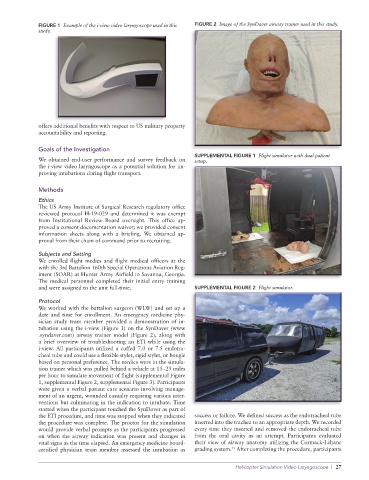
FIGURE 1  Example of the i-view video laryngoscope used in this   FIGURE 2  Image of the SynDaver airway trainer used in this study.
              study.


              offers additional benefits with respect to US military property
              accountability and reporting.


              Goals of the Investigation
                                                                 SUPPLEMENTAL FIGURE 1  Flight simulator with dual patient
              We obtained end-user performance and survey feedback on   setup.
              the i-view video laryngoscope as a potential solution for im-
              proving intubations during flight transport.

              Methods
              Ethics
              The US Army Institute of Surgical Research regulatory office
              reviewed protocol H-19-029 and determined it was exempt
              from Institutional Review Board oversight. This office ap-
              proved a consent documentation waiver; we provided consent
              information sheets along with a briefing. We obtained ap-
              proval from their chain of command prior to recruiting.

              Subjects and Setting
              We enrolled flight medics and flight medical officers at the
              with the 3rd Battalion 160th Special Operations Aviation Reg-
              iment (SOAR) at Hunter Army Airfield in Savanna, Georgia.
              The medical personnel completed their initial entry training
              and were assigned to the unit full-time.           SUPPLEMENTAL FIGURE 2  Flight simulator.

              Protocol
              We worked with the battalion surgeon (WLW) and set up a
              date and time for enrollment. An emergency medicine phy-
              sician study team member  provided a demonstration of in-
              tubation using the i-view (Figure 1) on the SynDaver (www
              .syndaver.com) airway trainer model (Figure 2), along with
              a brief overview of troubleshooting an ETI while using the
              i-view. All participants utilized a cuffed 7.0 or 7.5 endotra-
              cheal tube and could use a flexible stylet, rigid stylet, or bougie
              based on personal preference. The medics were in the simula-
              tion trainer which was pulled behind a vehicle at 15–25 miles
              per hour to simulate movement of flight (supplemental Figure
              1, supplemental Figure 2, supplemental Figure 3). Participants
              were given a verbal patient care scenario involving manage-
              ment of an urgent, wounded casualty requiring various inter-
              ventions but culminating in the indication to intubate. Time
              started when the participant touched the SynDaver as part of
              the ETI procedure, and time was stopped when they indicated   success or failure. We defined success as the endotracheal tube
              the procedure was complete. The proctor for the simulation   inserted into the trachea to an appropriate depth. We recorded
              would provide verbal prompts as the participants progressed   every time they inserted and removed the endotracheal tube
              on when the airway indication was present and changes in   from the oral cavity as an attempt. Participants evaluated
              vital signs as the time elapsed. An emergency medicine board-   their view of airway anatomy utilizing the Cormack-Lehane
                                                                             11
              certified physician team member assessed the intubation as   grading system.  After completing the procedure, participants
                                                                              Helicopter Simulation Video Laryngoscope  |  27