
Page 74 - JSOM Fall 2018
P. 74
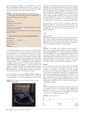
Study participants completed a short questionnaire to deter application. The radial pulse was used to assess LOS in the up
mine study eligibility, including exclusion criteria (Figure 1). A per limbs; the posterior tibial pulse was used to access LOS in
total of 41 volunteers were included in the study. The median the lower limb. The TQ was then activated by either inflating
age was 28 (range, 22–48) years and 68% were male. the cuff (pneumatic TQ), twisting the windlass rod (CAT),
or stretching and wrapping (SWATT). The TQ was left on as
FIGURE 1 Study participants completed a short questionnaire. long as necessary to record the pressure in the cuff, number of
Please circle any of the below if you have a history of: twists needed, or length of SWATT used to reach LOS, which
Myocardial infarction (heart attack) was approximately 30 seconds. The participant was allotted
2 minutes between each application to provide adequate time
Stroke
for reperfusion of the limb before continuing the experiment.
Heart surgery
Congenital vascular defects We performed t tests and χ tests to determine statistically
2
Hypertension significant differences between measured variables. Pain and
Trauma to an extremity that required fasciotomy or surgery pressure differences among anatomic comparisons were made
using the Wilcoxon signedrank test. A p value ≤.05 was con
necessitating hospital stay sidered significant.
Any comments or other health concerns you wish to reveal,
please list below:
LOS
Please write your answers in the underlined portion: Pneumatic TQ
What is your: When using the pneumatic TQ, one examiner would activate
the TQ by increasing the pressure with digital dials while an
Sex: Male ______ Female ______ other examiner would assess Doppler signal. When using the
Age (years): ______ SWATT or CAT, one examiner would apply and adjust the
Height (inches): ______ TQs while the other would assess Doppler signal.
Weight (lb): ______
C-A-T
With the CAT, which uses a windlass rod, the number of
To investigate whether pain may be used to predict TQ effi twists was recorded to determine the LOS. Each application
cacy, we sought to obtain pain values once TQs were deemed began with placing the CAT snug on the limb (one location
successfully placed. Three different TQ systems were used: a and TQ application at a time), placing the TQ mid biceps, mid
18.0 inch by 5.5 inch or 24.0 inch by 5.5 inch Stryker Single forearm, mid thigh, or mid calf. The first 90° rotation of the
®
Belly Pneumatic Tourniquet (Sustainability Solutions, http:// windlass placed the windlass parallel to the strap and was con
sustainability.stryker.com); 10.4cm elastic StretchWrapAnd sidered the baseline. From this point, the TQ was activate by
Tuck Tourniquet (SWATT ; H&H Medical Corp, www.swat twisting the windlass rod. The number of turns was recorded
™
tourniquet.com); and a 3.8cmwide Combat Application once LOS was reached. One twist was equivalent to 360°.
Tourniquet Generation 6 (CAT ; CAT Resources Inc, www
®
.combattourniquet.com) (Figure 2). TQs were applied to six SWAT-T
anatomic locations in random fashion: right upper arm (RUA), To accommodate for differences in limb size when the SWATT
right forearm (RFA), left upper arm (LUA), left forearm (LFA), (Figure 2) was used, the circumference of each limb was first
right thigh (RTH), and right calf (RCA). The circumferences measured in centimeters. The SWATT was then wrapped
of each of these sites were recorded in centimeters. around the limb just once, without applying any compressing
force. The length of the SWATT that was not wrapped around
For each TQ system, we used a 4MHz handheld Doppler ul the limb was considered the starting length. The SWATT was
trasound (MedLine, https://www.medline.com) to find LOS then activated by stretching and wrapping the TQ around the
and perceived this as loss of arterial pulse due to successful TQ limb until LOS was reached. The remaining length of unused
SWATT was measured and this was subtracted from the start
FIGURE 2 Stretch Wrap-And-Tuck Tourniquet and the Combat ing length to determine how much TQ was actually used to
Application Tourniquet. occlude the vasculature.
Pain
With each TQ model, at each anatomic location, after the TQ
was activated and LOS was appreciated with Doppler, sub
jects rated their pain on a 0 to 10 Visual Analog Pain Scale (0
being no pain, 10 being the worst pain; Figure 3). TQs were
removed once the pain rating was obtained.
FIGURE 3 Numeric pain scale.
0 1 2 3 4 5 6 7 8 9 10
No Moderate Worst
pain pain possible
pain
72 | JSOM Volume 18, Edition 3 / Fall 2018