
Page 134 - JSOM Spring 2020
P. 134
use of oral contraceptives and/or hormone replacement ther- if AT is not present) of each test. In both studies 48,49 ultrasound
apy. Oral contraceptives have been shown to decrease the was used to identify a “true” AT case and the ultrasound crite-
36
rate of collagen synthesis, at least partly explaining the latter ria were identical in both investigations. Note in Table 1 that
association. Biomechanically, prospective studies have found the within-tester reproducibility (intratester kappa coefficient)
39
risk factors include reduced or excessive ankle dorsiflexion can differ considerably, as indicated by the results of the study
range of motion, 7,40,41 less displacement of the center of force conducted by Maffulli et al., which included three clinicians.
48
during running, and a more laterally directed force distribution This suggests that clinicians need to train on these tests as
when the foot is flat during running. At least two systematic was done in the study of Hutchison et al., where generally
42
49
reviews have indicated that consumption of antibiotics in the higher reproducibility was achieved. Table 1 suggests that on
fluoroquinolone class substantially increases AT risk 43,44 with the basis of accuracy and reproducibility, the morning stiff-
one meta-analysis indicating a 4-fold risk increase (odds ratio ness, self-reported pain, palpation, and tendon thickening tests
= 3.95, 95% confidence interval (95% CI) = 3.11–5.01). 44 appear to be the most useful, although false positives were
high on the morning stiffness test, and there was considerable
The DMED provides data that can be used to examine a lim- variability between the two studies 48,49 on the sensitivity of the
ited number of demographic factors that might be associated palpation test.
with AT in military personnel. To examine these potential
risk factors DMED data on the incidence of AT (ICD-9 code If the clinical evaluation is inconclusive imaging studies can
726.71) was compiled by sex, age, race, and military service be useful. Both ultrasound and magnetic resonance imaging
for the years 2001 to 2015. Incidence was calculated using the (MRI) have been used in this regard. Ultrasonography is read-
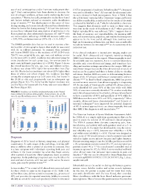
entire population for each group (e.g., for women [new fe- ily available and less expensive, but it is operator dependent,
male cases (n)/female population (n) × 1000]). Figure 5 shows provides only a two-dimensional image, and transducer han-
the overall incidence by sex, age, race, and military service. dling and machine settings can influence the images. MRI pro-
Incidence was about 10% higher for women than men (Fig- vides better images because the three-dimensional structure of
ure 5A). Blacks had an incidence rate about 22% higher than tendon can be acquired and there is better ability to visualize
those of whites and others (Figure 5B). Incidence was high soft tissue. Further, MRI can assist in differentiating between
among the youngest age group (<20 years old), but lowest in stages of the AT process and between paratenonitis and ten-
the 20-24 yr olds and progressively rose in subsequent age dinosis. 24,45,47 In head-to-head comparisons, MRI has proven
groups (Figure 5C). The Army had the highest incidence rate somewhat more accurate than ultrasonography in diagnosing
and the Navy the lowest with the Army rate over twice that of AT. In one study of histologically verified AT, ultrasound cor-
the Navy (Figure 5D). rectly identified AT cases 81% of the time while with MRI
96% of cases were correctly identified. In another study that
50
FIGURE 5 Incidence of Achilles tendinitis/bursitis in the United used clinical assessments as the criterion, ultrasound was found
States military by various demographic characteristics. A, sex; B,
race; C, age; D, military service. MC, Marine Corps, AF, Air Force. to have a sensitivity of 80% and specificity of 49% while for
MRI these numbers were 95% and 50%, respectively. More
51
recently, ultrasound tissue characterization and B-mode ul-
52
trasound techniques have improved the potential diagnosis
53
of AT. A rational approach is to use ultrasonography first and
then MRI if the diagnosis is still inconclusive. 47,54
The Victorian Institute of Sports Medicine Assessment–Achil-
les (VISA-A) is a simple eight-item questionnaire that can be
used to assess the severity of AT and track clinical progress.
The VISA-A assesses three domains comprising pain, func-
tional status, and activity with each question rated on a 5- to
10-point scale. In the study that developed the test, the test-re-
test reliability was r = 0.93, between-rater reliability r = 0.90,
and within-rater reliability r = 0.90. The test was designed so
that a score of 100 would indicate a totally asymptomatic indi-
Diagnosis
vidual so higher scores indicate less pain and higher function.
Diagnosis of AT is based on history, symptoms, and physical Patients awaiting surgical care for Achilles problems scored 44
examination. In early stages of the pathology, morning stiff- ± 28, symptomatic patients scored 64 ± 17, and asymptomatic
ness or stiffness after a period of inactivity is common. Pain patients scored 96 ± 7. In a randomized controlled trial of
55
is a later symptom with individuals reporting both pain and treatment options for AT, the VISA-A scores tracked well with
stiffness in the lower posterior leg when they begin activity other clinical tests that indicated improvements in pain and
after a period of inactivity. These symptoms lessen as activity function. The test can be obtained at https://bjsm.bmj.com/
56
progresses. As the pathology progresses pain may be felt at the content/suppl/2001/11/09/35.5.335.DC1/01055_Fig_1_data_
start and end of activity with less pain while active. In severe supplement.pdf.
cases, pain may be present at rest. 45–47
Silverskold test can be useful to distinguish between gastroc-
In two studies, 48,49 to a total of 10 clinical tests were evaluated nemius versus Achilles tendon and soleus muscle tightness.
for their ability to assist in the diagnosis of AT. Table 1 de- In this test, the patient is supine and the tester first evalu-
scribes these tests, defines a positive outcome (i.e., presence of ates ankle dorsiflexion with the knee flexed, then extended.
AT), and provides the reported sensitivity (ability to correctly More dorsiflexion with the knee flexed indicates gastrocne-
identify an actual AT case) and specificity (ability to determine mius tightness. This is because the gastrocnemius becomes less
128 | JSOM Volume 20, Edition 1 / Spring 2020