
Page 132 - JSOM Spring 2020
P. 132
Terminology is important when focusing on AT because many
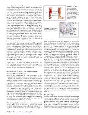
nomenclatures have been used to describe tendon pathologies FIGURE 1 Simplified
and the use of terms has evolved over time as the disorder has macro-structure of
the Achilles tendon
become better understood. In the past, tendinitis was a general showing major muscle
term used to describe pain and/or swelling of a tendon while groups and attachment
implying inflammatory processes. The term tendinosis was of Achilles tendon to
later suggested to imply tendon degradation, while remov- the calcaneus (modified
from https://www.
ing the focus from inflammation since there was debate over physio-pedia.com/
whether inflammation, in the traditional sense, was present Achilles_Rupture).
within the tendon structure. Paratenonitis (also called periten-
dinitis, tenosynovitis, and tenovaginitis) referred to the inflam-
mation of the connective tissue surrounding the tendon. 1,2,9–11
It was often difficult clinically to distinguish between these
disorders because the presentation could be very similar and
concurrent. Most recently, several practitioners have advo- FIGURE 2 Achilles tendon
cated that the term AT be used. AT is a general descriptor for a micro-structure (from 17
nonrupture injury of the tendon, paratenon, or both in which Towler and Gelberman ).
the patient presents with pain, swelling, and reduced perfor-
mance and for which symptoms are exacerbated by excessive
mechanical loading due to physical activity. 1–3,12 It has been
suggested that the other terms only be used when there is or is
1
not histological evidence of inflammatory cells.
within it and the paratenon that surrounds it, contains the
Some clinicians 3,13 have also advocated that AT be separated blood vessels, lymphatics, and the nerve supply of the tendon.
into two different clinical entities: insertional AT and midpor- Fiber bundles contain tenocytes, collagen fibers, and ground
tion AT. The difference is based on anatomic location, symp- substance. The tenocytes are cells within the tendons that
toms, and histopathological findings. Insertional AT involves maintain all the components of the extracellular matrix. Teno-
pain stiffness and often swelling at the insertion of the Achilles cytes respond to mechanical stimulation (e.g., exercise) by
tendon into the calcaneus. Bone spurs and calcification may increasing collagen formation and producing other proteins.
be present and palpable at the insertion and small tears of the Collagen fibers are arranged primarily longitudinally along
tendon tissue may be present. In midportion AT there is pain the long axis of the tendon, although some fibers can run in
with diffuse or localized swelling 2cm to 7cm superior to the other directions. There are at least 28 different collagen pro-
calcaneal insertion with disruption of the tendon fibers and teins, but about 90% of the collagen in the tendon is type 1,
increased tendon vascularity. which has very high tensile strength, allowing it to withstand
high forces from both ends of the tendon. Ground substance
The purpose of this article is to provide an overview of the is composed of several molecules among the most important
pathophysiology, epidemiology, diagnosis, treatment, preven- of which are proteoglycans. Proteoglycans attract water, assist
tion, and prediction of AT. Where possible, a distinction will in maintaining the tendon, and provide collagen fibers with
be made between insertional and midportion AT. structural support. Water is 65% to 75% of the weight of the
tendon and water with the proteoglycans provide lubrication
Achilles Tendon Structure and Pathophysiology and spacing for the gliding function of the tendon. The tertiary
fiber bundles contain secondary and primary fiber bundles
Structure of the Achilles Tendon (also called fascicles and subfascicles, respectively) that con-
Before the pathophysiology of AT can be appreciated it is nec- tain collagen fibrils. All primary, secondary and tertiary fiber
essary to understand the macro- and micro-structure of the bundles are encased in the connective tissue network called
Achilles tendon. Tendons are soft tissue structures connecting endotenon. The tendon fiber bundles themselves are not well
muscle to bone and allowing for the transmission of forces from supported with blood vessels with these instead primarily ly-
muscular contraction to the bone to produce movement. The ing between the tertiary fiber bundles in the endotenon. 16–21
Achilles tendon is the longest, largest, and strongest tendon in Finally, the whole tendon complex, encased by the epitenon,
the body. 14,15 As shown in Figure 1, the Achilles tendon connects is surrounded by a loose layer of areolar connective tissue that
the two-headed gastrocnemius and single-bodied soleus muscles comprises the paratenon—the role of the paratenon is to re-
to the calcaneus to produce ankle plantar flexion, a very import- duce friction between the tendon as a whole and surround-
ant movement for running and walking. The Achilles tendon in- ing tissues as the tendon moves relative to these surrounding
serts medially and laterally on the calcaneus forming a crescent tissues. 12
shape that transmits muscular forces while distributing the force
across a large portion of the calcaneus. Two bursae surround Pathophysiology
the tendon near its insertion at the calcaneus: the subcutaneous Repetitive and excessive loading of the Achilles tendon during
bursa is between the skin and tendon while the retrocalcaneal physical activity (e.g., running, jumping) appears to be the
bursa is between the tendon and calcaneus. 15 main stimulus for tendinopathy. 22,23 In overuse tendinopathy,
frequent cumulative microtrauma can cause tendon damage
The well-organized micro-structure of the tendon is shown in that is adaptive (can be repaired) or nonadaptive (causing de-
Figure 2. The epitenon is composed of a thin sheet of connec- generation of the tendon). It has been proposed 24,25 that ten-
tive tissue and surrounds the tendon. It, along with the loose don pathology due to overloads experienced during repetitive
endotenon layers that bind the tendon fiber bundles together physical activity can be viewed as a continuum of changes. For
126 | JSOM Volume 20, Edition 1 / Spring 2020