
Page 100 - JSOM Winter 2018
P. 100
application of tourniquets when compared with traditional Material, https://www.dickblick.com/,) with salt and water.
instructional models alone. A secondary aim of this study was While nonpulsatile, this method allows tourniquet placement
to evaluate the confidence of the trainees in their self-reported without tubing rupture associated with positive displacement
understanding of the indications for and technical abilities to pumps.
apply tourniquets to exsanguinating limb injury after training.
After the instruction and practice sessions (TT or PCT), each
corpsman was brought to an unmarked and covered cadaver
Methods
(with hospital gown and sheet). Once the gown and sheet
This study was performed after approval by the Institutional were removed (to simulate injury and wound exposure) and
Review Board at the Keck School of Medicine of USC and in extremity hemorrhage was identified, each of the corpsmen
accordance with the Keck School of Medicine of USC Fresh performed tourniquet application on the right and left lower
Tissue Dissection Laboratory (FTDL) policies. From Janu- extremity in separate timed events. The time taken to place the
ary 2016 to November 2016, US Navy corpsmen rotating at tourniquet(s) and stop the bleeding was recorded. After the
NTTC were recruited to participate. Fifty-three corpsmen vol- tourniquet(s) was/were secured, a trauma surgeon blinded to
unteered for the study. Demographic data were collected and the teaching method assessed the position of the tourniquet(s).
included age, sex, experience (years), deployment history, and
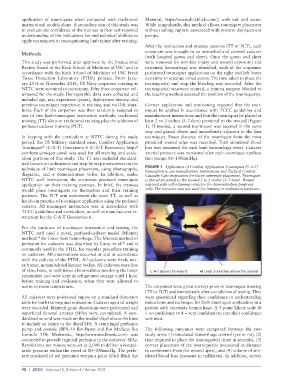
previous tourniquet experience in training and real-life situa- Correct application and positioning required that the tour-
tions. Each of the corpsmen was then randomly assigned to niquet be applied in accordance with TCCC guidelines and
one of two limb-tourniquet instruction methods: traditional manufacturer instructions and that the tourniquet be placed at
training (TT) alone or traditional training plus the addition of least 2 to 3 inches (5–7.6cm) proximal to the wound (Figure
perfused-cadaver training (PCT). 1). If needed, a second tourniquet was secured in the same
way and placed above and immediately adjacent to the first
In keeping with the curriculum at NTTC during the study tourniquet. Exact distance of the tourniquet from the most
period, the US Military standard-issue, Combat Application proximal wound edge was recorded. Total simulated blood
Tourniquet (C-A-T) Generation 6 (C-A-T Resources; http:// loss was measured for each limb hemorrhage event. Cadaver
®
combattourniquet.com/) was used for all training and evalu- arterial pressure was measured after each tourniquet applica-
ation portions of this study. The TT arm included standard- tion (range, 80–100mmHg).
ized lecture on indications and step-by-step instructions on the
technique of limb tourniquet placement, using photographs, FIGURE 1 Application of Combat Application Tourniquet (C-A-T)
diagrams, and a demonstration video. In addition, under Generation 6, per manufacturer instructions and Tactical Combat
Casualty Care instruction for lower extremity placement. Tourniquet
NTTC staff instruction, the corpsmen practiced tourniquet is placed proximal to the wound 2 to 3 inches (5–7.6cm). Wound
application on their training partners. In brief, the trainees exposed with self-retaining retractor for demonstration purposes
would place tourniquets on themselves and their training only. The retractor was not used for training or evaluation purposes.
partners. The PCT arm underwent the same TT, as well as
hands-on practice of tourniquet application using the perfused
cadaver. All tourniquet instruction was in accordance with
TCCC guidelines and curriculum, as well as manufacturer in-
struction for the C-A-T Generation 6.
For the purposes of tourniquet instruction and testing, the
NTTC staff used a novel, perfused-cadaver model (Minneti
method) for lower limb hemorrhage. The Minneti method of
10
perfusion for cadavers was described by Carey et al. and is
10
commonly used in the FTDL for vascular procedure training
on cadavers. All interventions occurred at and in accordance
with the policies of the FTDL. All cadavers were fresh, nev-
er-frozen, nonembalmed human bodies. All cadavers were free
of skin, bone, or soft-tissue abnormalities involving the lower
extremities and were kept in refrigerated storage until 1 hour
before training and evaluation, when they were allowed to
warm to room temperature. The corpsmen were given surveys prior to tourniquet training
(TT or PCT) and immediately after completion of testing. They
All cadavers were positioned supine on a standard dissection were questioned regarding their confidence in understanding
table for both training and evaluation. Cadaver age and weight indications and technique for limb tourniquet application in a
were recorded. Bilateral groin dissections were performed and patient with extremity hemorrhage. A 5-point Likert scale (0
superficial femoral arteries (SFAs) were cannulated. A stan- = no confidence to 4 = very confident) to rate their confidence
dardized wound was made on the medial thigh above the knee was used.
to include an injury to the distal SFA. A centrifugal perfusion
pump and console (BPX-50 Bio-Pump and Bio Medicus Bio The following outcomes were compared between the two
Console 550; Medtronic, http://www.medtronic.com) was study arms: (1) simulated hemorrhage control (yes or no), (2)
connected to provide regional perfusion in the cadavers’ SFAs. time required to place the tourniquet(s) (time in seconds), (3)
Revolutions per minute were set at 2,000 to deliver a nonpul- correct placement of the tourniquet(s) (measured as distance
satile pressure within the vessel of 80–100mmHg. The perfu- in centimeters from the wound apex), and (4) volume of sim-
sate consisted of red premium tempura paint (Dick Blick Art ulated blood loss (measure in milliliters). In addition, survey
98 | JSOM Volume 18, Edition 4 / Winter 2018