
Page 53 - JSOM Fall 2018
P. 53
and Department of Defense regulations. Animals were main alarm signals due to overpressure. The Belmont is designed to
tained in a facility accredited by the Association for Assessment sound an alarm, display a “High Pressure” message, and stop
and Accreditation of Laboratory Animal Care International in the transfusion at the factorydetermined maximum pressure
accordance with the Guide for the Care and Use of Laboratory limit at or above 300mmHg. When the highpressure alarm
Animals. Our study included nine Yorkshire swine (Sus scrofa), sounded, the research assistant silenced the machine and man
18
weighing between 70kg and 90kg. This weight range was chosen ually restarted the transfusion. The number of pressure alarms
because it represents the 50th percentile bone density range of the during a 5minute interval was recorded.
average adult male 20 to 39 years old, the group that constitutes
the majority of our Combat Forces. 12,19 All animals were healthy, Push-pull transfusion
intact females. No animals were excluded because of disease, in In the manual pushpull arm, three swine were transfused with
jury, or illness before commencement of the study. Females were either a 10mL, 20mL, or 60mL syringe connected to a three
chosen because prior research reported minimal differences in way stopcock. The 50mL syringe transfusion method was pre
bone density among male, female, and barrow swine. 20 viously described by the British Medical Emergency Response
Team during Operation Enduring Freedom. Pediatric resus
22
Transfusion Strategies citation literature advocates for 10mL or 20mL syringes to
Animals were randomly assigned to one of the following four decrease hand fatigue. 23
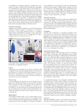
transfusion strategies (Figure 1): (1) gravity (n = 2); (2) pres
sure bag (n = 2); (3) rapidtransfusion device (n = 2; Belmont Anesthesia
Rapid Infuser pump; Belmont Instrument Corp., http://www Activities were conducted in a controlled, designated veteri
.belmontinstrument.com/); and (4) manual pushpull using a nary surgical suite. Animals were fasted before anesthesia and
syringe and threeway stopcock (n = 3). 1,21,22 premedicated with glycopyrolate (0.05mg/kg intramuscularly
[IM]), ketamine (20mg/kg), and xylazine (2mg/kg). Once endo
FIGURE 1 Intraosseous transfusion strategies. tracheal intubation was achieved, the animals were mechani
cally ventilated with a mixture of isoflurane and oxygen, using
a large animal veterinary anesthesia machine (Hallowell EMC,
www.hallowell.com/). Butorphanol (0.2mg/kg IM) was given
for analgesia. Vital signs were continuously monitored every
15 minutes. All animals were euthanized at the completion of
the protocol under general anesthesia.
Experimental Hemorrhage
To establish baseline clotting strength, three thromboelasto
gram (TEG) measurements were performed for each animal
before inducing hemorrhage. In addition, three TEG measure
ments were performed at the conclusion of transfusion, and at
time of death or 1hour after transfusion, whichever occurred
first. The left carotid artery was exposed using a standard cut
down technique and cannulated using a Seldinger technique
Left to right: Gravity transfusion; pressure bag transfusion; Belmont with a 9F introducer catheter for arterial, invasive blood
Rapid Infuser; and pushpull transfusion. pressure monitoring during the experimental procedures. Us
ing a similar cutdown technique, the femoral artery was can
Gravity nulated with a 9F introducer catheter to induce a controlled
In the gravity arm, transfusion in two swine occurred through hemorrhage. After instrumentation, there was a 10minute
a single IO access in the left proximal humerus via the pressure stabilization period during which baseline hemodynamic data,
generated from the weight of the blood hanging from a stan including hemoglobin, hematocrit, an electrolyte panel, and
dard pole for hanging intravenous (IV) fluids and medications lactate were collected. These values were repeated immediately
The IV pole was positioned lateral to the proximal humerus after transfusion and at time of death or 1hour after transfu
IO insertion site prior to transfusion. sion, whichever occurred first.
Pressure bag A controlled hemorrhage was performed by collecting blood
In the pressurebag arm, the first animal had a single IO access through the force of gravity into a 450mL bloodbag system
placed in the left proximal humerus, a bag of blood was hung containing 63mg of citrate phosphate dextrose adenine. The
from a standard IV pole, and a pressure bag was inflated to bloodcontainer bag was located on a weight scale and rocker
and maintained at or above 300mmHg by a member of the (Genesis BPS, http://www.genesisbps.com/) below the animal.
research team. The second animal in the pressurebag arm had An intended range of 20–25mL/kg blood was removed over 15
IO access placed in the left and the right proximal humeri and to 40 minutes. Hemorrhage and blood collection were halted
blood was transfused simultaneously through both sites using if the mean arterial pressure (MAP) reached 30mmHg or less.
a pressure bag. After experimental hemorrhage, specimens were collected for
testing and analysis. After hemorrhage completion, each animal
Rapid infusion was given 30 minutes to allow time for vital signs to stabilize.
In the Belmont Rapid Infuser arm, the rapid infuser was set
up according to manufacturer guidelines. The transfusion rate IO Access
was set at 100mL/min in the first animal and 50mL/min in the After the stabilization period, an IO catheter was placed ac
second animal to detect any difference in frequency of machine cording to the manufacturer’s instructions. A 15gauge, 45mm,
Four Intraosseous Blood Transfusion Strategies | 51