
Page 89 - JSOM Summer 2018
P. 89
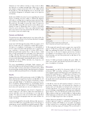
Operator can heal without making an injury worse or allow   TABLE 1  MRI Frequency
              the Operator to continue training after ruling out an injury   Site               Frequency, n
              that requires surgical intervention, PT, or rest. MRIs should   Knee                 10
              be included as a first-line diagnostic tool to aid in the rapid   Shoulder            9
              and definitive diagnosis of orthopedic injury in the Operator
              community.                                         Elbow                              3
                                                                 Lumbar spine                       3
              Earlier use of MRI over plane X-rays is appealing for several   Cervical spine        3
              reasons, including increased ability to definitively diagnose   Ankle                 3
              soft-tissue or cartilage injury requiring surgery, increased mis-  Wrist             2
              sion readiness, and elimination of unnecessary exposure to IR.   Tibia and fibula    1
              We undertook this study to review the results of experience   Foot                   1
              with early MRI among PJs and combat rescue officers com-
              plaining of pain or dysfunction due to injury. A secondary   TABLE 2  Diagnosis by Injury Type
              objective was to determine if there was a high positivity rate
              due to the nature of the men selected for this work (i.e., high   Injury Type     Frequency, n
              thresholds of pain and complaining).               Tendon                             8
                                                                 Other soft tissue                  7 a
                                                                 Bone                               7
              Patients and Methods                               Cartilage                          6
              This performance improvement project was approved by the   Disc                       6
              institutional review board of the Air Force Research Oversight   Ligament             5
              and Compliance Division.                           Arthropathy                        5 b
                                                                 a Bursitis, fasciitis, muscle tear, popliteal cyst.
              From April 2008 through December 2014, the injuries of 45   b Bone contusions, periosteal reaction, bone cyst.
              PJs on a single team were managed by a single flight surgeon.
              On  injury or  complaint  of  worsening  of an  intermittent  or   18 PJs examined required surgery; surgery was required for
              chronic injury associated with significant pain or dysfunction   eight of 15 PJs if the lumbar spine MRIs are excluded (spine
              (e.g.,  weakness  or  decrease  range  of  motion),  MRI  was  or-  MRI was performed for pain in the absence of weakness or
              dered and performed anytime from 1 day to about 1 week   sensory loss). In all cases of surgical intervention, treatment
              later. A single musculoskeletal radiologist (C.F.), who became   was definitive. In nonsurgical cases, PT, active recovery, con-
              familiar with the Operators and the job demands, read ev-  tinued training, and rest were prescribed on the basis of the
              ery MRI. Records were maintained prospectively and collated   exact diagnoses.
              retrospectively. Information regarding the indication for MRI,
              mechanism of injury (MOI), anatomic site, diagnoses, and   Of the 35 MRIs performed, including the spine MRIs, 21
              treatment was recorded.                            (60%) were positive and directed Operators for further care,
                                                                 rest, or resumption of training.
              The same musculoskeletal radiologist, flight surgeon, and
              sports fellowship–trained orthopedic surgeon reviewed all re-
              sults and, when necessary, discussed the treatment plan, which   Discussion
              incorporated the operational and clinical perspectives. In most   Approximately one-half of the Operators (eight of 15) who
              instances, MRI was performed before orthopedic consultation.  underwent MRI (excluding lumbosacral studies) over 6.5
                                                                 years required surgical intervention. In a small squadron com-
              Results                                            posed of 45 total Operators, 35 MRIs were performed on 18
                                                                 individuals (40%), of whom eight (17.7% of the total Oper-
              Eighteen Operators (40%) underwent a total of 35 MRIs. The   ators on the team) required surgical intervention. Therefore,
              most common sites imaged were the knee and shoulder (n = 10   during the observed time period, almost one in five Operators
              and 9, respectively), followed by elbow, cervical spine, lumbar   underwent surgery.
              spine, and ankle (Table 1). The MOI was categorized as over-
              use in 16 patients, military training (e.g., parachuting, adverse   It should be noted that all surgeries resulted in clinical im-
              terrain training, battle drills) in 11, acute physical training in-  provement. Surgeries were not performed for diagnosis of
              jury in six, acute sports injury in one, and unknown in one.  injuries such as small labral or meniscal tears not felt to be
                                                                 clinically significant. This may be a reflection of the team ap-
              The most common diagnoses were related to ligament and   proach including a musculoskeletal radiologist and orthopedic
              tendon injuries (n = 18; Table 2). There were three bone con-  sports surgeon, who have developed a good rapport with the
              tusions, and there were disc protrusions on the four spine   flight surgeon and the team, which includes an understanding
              studies. The only pure bone abnormalities that would have   of the Operators and their job and the notion that they do not
              shown up on chest radiographs were two cases of distal cla-  come to the flight surgeon for minor complaints.
              vicular osteolysis.
                                                                 If these Operators presented to a general medical group, the
              Treatment was guided by the results. Eleven of the injuries re-  standard approach would be conventional radiograph fol-
              viewed with MRI were treated with surgical intervention; no   lowed by 6 weeks of PT. If this is not successful, the patients
              spine injuries viewed with MRI were treated surgically. There-  are then referred for an orthopedic consultation, then MRI,
              fore, 11 of the injuries viewed with MRI among eight of the   and then scheduling surgery, if indicated. This would result in

                                                                        MRI in Optimizing Injury Management in Operators  |  87