
Page 29 - JSOM Spring 2018
P. 29
A portable chest radiograph showed the ETT was in the tra- edema secondary to volume overload. The patient tolerated
chea approximately 10cm above the carina with no evidence a spontaneous ventilator mode with Fio of 40%, pressure
2
of acute cardiopulmonary abnormalities. The ventilator respi- support of 10cmH O, and PEEP of 5cmH O. He maintained
2
2
ratory rate was increased to 18/min to reduce the respiratory stable Sao at 99%–100%, with RR of 18–20 bpm.
2
acidosis. Deep tracheal suctioning was performed and the ETT
was advanced 2cm. During this intervention, telemetry showed The norepinephrine bitartrate drip was stopped at 01:00, 11
a 6-beat run of ventricular tachycardia then an 8-beat run fol- hours after ICU admission. The bicarbonate drip was stopped
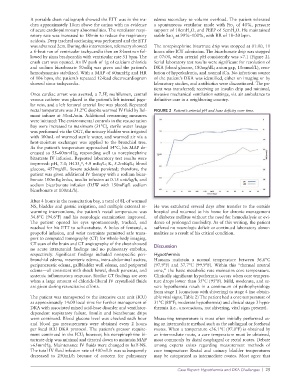
lowed by sinus bradycardia with ventricular rate 51 bpm. The at 07:00, when arterial pH consistently was >7.1 (Figure 2).
crash cart was opened. An IV push of 1g of calcium chloride Serial laboratory test results were significant for resolution of
and sodium bicarbonate 50mEq was given and the patient’s DKA (blood glucose, 180mg/dL; anion gap, 10mmol/L), reso-
hemodynamics stabilized. With a MAP of 60mmHg and HR lution of hyperkalemia, and normal iCa. No infectious source
of 106 bpm, the patient’s repeated 12-lead electrocardiogram of the patient’s DKA was identified, either on imaging or by
showed sinus tachycardia. laboratory studies, and antibiotics were discontinued. The pa-
tient was transferred; receiving an insulin drip and minimal,
Once cardiac arrest was averted, a 7.5F, multilumen, central invasive mechanical ventilation settings, via air ambulance to
venous catheter was placed in the patient’s left internal jugu- definitive care in a neighboring country.
lar vein, and a left femoral arterial line was placed. Repeated
rectal temperature was 31.2°C despite warmed IV fluid by Bel- FIGURE 2 Patient’s arterial pH and base deficity over time.
mont infuser at 50mL/min. Additional rewarming measures
were initiated: The environmental controls in the resuscitation
bay were increased to maximum (31°C), sterile water lavage
was performed via the OGT, the urinary bladder was irrigated
with 300mL of warmed sterile water, and warmed air via a
heat-moisture exchanger was applied to the bronchial tree.
As the patient’s temperature approached 34°C, his MAP de-
creased to 55–60mmHg, responding well to norepinephrine
bitartrate IV infusion. Repeated laboratory test results were
improved: pH, 7.0; HCO − , 4.9 mEq/L; K, 5.2mEq/L; blood
3
glucose, 457mg/dL. Severe acidosis persisted; therefore, the
patient was given additional IV therapy with a sodium bicar-
bonate 100mEq bolus, insulin infusion at 0.15 unit/kg/h, and
sodium bicarbonate infusion (D5W with 150mEq/L sodium
bicarbonate at 100mL/h).
After 4 hours in the resuscitation bay, a total of 8L of warmed
NS, bladder and gastric irrigation, and multiple external re- He was extubated several days after transfer to the outside
warming interventions, the patient’s rectal temperature was hospital and returned to his home for chronic management
34.8°C (94.6°F) and his neurologic examination improved. of diabetes mellitus without the need for hemodialysis or evi-
The patient opened his eyes spontaneously, tracked, and dence of prolonged morbidity. As of this writing, the patient
reached for his ETT to self-extubate. A bolus of fentanyl, a suffered no neurologic deficit or continued laboratory abnor-
propofol infusion, and wrist restraints permitted safe trans- malities as a result of his critical condition.
port to computed tomography (CT) for whole-body imaging.
CT scan of the brain and CT angiography of the chest showed Discussion
no acute intracranial findings and no pulmonary embolus,
respectively. Significant findings included nonspecific peri- Hypothermia
bronchial edema, mesenteric edema, intra-abdominal ascites, Humans maintain a normal temperature between 36.6°C
peripancreatic edema, gallbladder wall edema, and periportal (97.9°F) and 37.7°C (99.9°F). Within this “thermal neutral
edema—all consistent with shock bowel, shock pancreas, and zone,” the basic metabolic rate maintains core temperature.
systemic inflammatory response. Similar CT findings are seen Clinically significant hypothermia occurs when core tempera-
when a large amount of chloride-liberal IV crystalloid fluids ture drops lower than 35°C (95°F). Mild, moderate, and se-
are given during resuscitative efforts. vere hypothermia result in a continuum of pathophysiology
from stage 1 (conscious with shivering) to stage 4 (no obtain-
The patient was transported to the intensive care unit (ICU) able vital signs; Table 2) The patient had a core temperature of
at approximately 14:00 local time for further management of 31°C (88°F; moderate hypothermia) and clinical stage 3 hypo-
DKA with associated mixed acid-base disorder and ventilator- thermia (i.e.. unconscious, not shivering, vital signs present).
dependent respiratory failure. Insulin and bicarbonate drips
were continued. Blood glucose level was checked each hour Measuring temperature is most often initially performed us-
and blood gas measurements were obtained every 2 hours ing an intermediate method such as the sublingual or forehead
per local ICU DKA protocol. The patient’s pressor require- routes. When a temperature <36.1°C (97.0°F) is obtained by
ment continued in the ICU; however, his norepinephrine bi- an intermediate route, a core temperature must be obtained,
tartrate drip was minimal and titrated down to maintain MAP most commonly by distal esophageal or rectal routes. Debate
>65mmHg. Maintenance IV fluids were changed to half-NS. among experts exists regarding measurement methods of
The total IV fluid infusion rate of 400mL/h was subsequently core temperature: Rectal and urinary bladder temperatures
decreased to 250mL/h because of concern for pulmonary may be categorized as intermediate routes. Most agree that
Case Report: Hypothermia and DKA Challenges | 25