
Page 28 - JSOM Spring 2018
P. 28
The decision was made to intubate, using etomidate for induc- of the warmed 1L NS bolus were given. The patient’s HR re-
tion and succinylcholine for paralysis. Despite three attempts sponded to these interventions. Telemetry showed normal si-
to intubate with direct laryngoscopy, the vocal cords were not nus rhythm of 95 bpm and MAP of 75mmHg; however, within
well visualized. The fourth attempt with a bougie adjunct was 5 minutes, his rhythm reverted to atrial fibrillation at a rate of
successful, and the patient proceeded at 07:00 local time with 130 bpm. An additional diltiazem 10mg IV bolus resulted in
the Dustoff crew to an alternate Role 2 facility, where a tail-to- normal sinus rhythm at 75 bpm and MAP of 60mmHg.
tail handoff was planned. A mechanical malfunction of the pa-
tient’s endotracheal tube (ETT) occurred en route to the Role 2 Initial arterial blood gas (ABG) sample results were as fol-
facility; the ETT endcap was dislodged. Dustoff Medics were lows: pH, 6.681; partial pressure of carbon dioxide (Pco ),
2
able to oxygenate the patient through a poorly seated bag- 42mmHg; partial pressure of oxygen (Po ), 145mmHg; base
2
valve-mask on the uncapped ETT, with tape used as a field- deficit, −30mEq; sodium (Na), 132mEq/L; potassium (K),
expedient connector. The FST performed a tube exchange in 6.4mEq/L; chloride (Cl), 111mEq/L; bicarbonate (HCO − ),
3
one successful attempt. During the procedure, the patient was <5mEq/L, ionized calcium (iCa), 1.06mmol/L, hemoglobin,
paralyzed with 10mg of vecuronium IV, sedated with 2mg of 11.9g/dL; and lactate, 2.8mmol/L, consistent with severe an-
versed IV, and given 100mg of ketamine IV. Once the airway ion-gap metabolic acidosis (AGMA), respiratory acidosis, and
and invasive mechanical ventilation were secured, the Dustoff nonanion-gap metabolic acidosis (NAGMA; Table 1). Initial
crew proceeded with the patient to the Role 3 facility, arriving diagnoses were moderately severe hypothermia, diabetic ke-
at 10:10 local time (almost 12 hours after initial medevac). toacidosis (DKA), and atrial fibrillation with rapid ventricular
response complicated by hypovolemic shock. The renal panel
In the resuscitation bay at the Role 3 facility, the patient was at T+40 minutes confirmed mild acute kidney injury (blood
on a litter in a Blizzard Rescue Blanket (Blizzard Protection urea nitrogen [BUN]/creatinine [Cr] ratio, 161.3), hyperkale-
Systems, http://www.blizzardsurvival.com) without a head mia, and hyperchloremic acidosis.
cover and lying supine on a cold Ready-Heat Blanket (Tech-
Trade, http://www.ready-heat.com/). On examination, he was
intubated with a 7.0F ETT, 19cm at the teeth. The Glasgow TABLE 1 Interpretation of Acid-Base Disorders With Brief
Discussion of Arterial Blood Gas Analysis
Coma Scale score was 3T; the patient had sluggishly reactive
bilateral pupils, was breathing passively on the ventilator set Arterial blood pH, 6.682; Pco , 42.3mmHg; Po ,
2
2
gas values
145mmHg; base excess, −30mEq; Sao , 94%
at assist control, fraction of inspired oxygen (Fio ) was 100%, 2
2
RR was 10/min, total lung volume was 450mL, and positive Basic metabolic Na, 132mEq/L; K, 6.4mEq/L; Cl, 110mEq/L;
−
HCO , <5mEq/L; BUN, 16mg/dL; Cr, 1.3mg/
panel
3
end-expiratory pressure (PEEP) was 5cmH O. Other invasive dL; blood glucose, 552mg/dL; anion gap,
2
devices included two peripheral intravenous lines, an oral-gas- 22mEq/L; lactate, 2.8mmol/L
tric tube (OGT), and a Foley catheter drain containing 150mL Step 1: Determine The patient has a primary metabolic acidosis,
−
of light yellow urine. The patient’s initial rectal temperature the primary acid- indicated by the low pH and low HCO level.
3
was 31°C (88°F), blood pressure was 153/94mmHg (mean ar- base disorder The patient’s acidosis is an AGMA, because
terial pressure [MAP], 92mmHg), HR was 165 bpm, RR was the anion gap is 22mEq/L (normal, 12mEq/L).
Given the mildly elevated lactate level, the
10/min, and Sao was 99% on 100% Fio . His examination cause of the patient’s AGMA is ketoacidosis.
2
2
was remarkable for tachycardia, obtundation, paralysis, and Step 2: Is the Because the patient’s primary disorder is
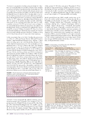
cold extremities. His 12-lead electrocardiogram demonstrated compensation AGMA, the appropriate compensation
atrial fibrillation with a ventricular rate of 161 bpm and for the primary is respiratory alkalosis. To determine the
prominent Osborn waves in the precordial leads (Figure 1). disorder expected Pco , use the formula Pco = 1.5 ×
2
2
−
appropriate? HCO + 8. In our patient, the expected Pco
2
3
FIGURE 1 Twelve-lead electrocardiogram demonstrating atrial for appropriately compensated AGMA is
fibrillation with a ventricular rate of 161 bpm and prominent 8mmHg. His Pco is 42mmHg, which means,
2
Osborn waves in the precordial leads. in addition to AGMA, he has severe respiratory
acidosis that likely is iatrogenic, because he was
paralyzed and, without any respiratory drive,
his minute ventilation was fully ventilator
dependent.
Step 3. Is there a Because the patient has AGMA, it is necessary
Δ gap? to rule out a mixed acid-base disorder such
as metabolic alkalosis or NAGMA. First,
determine the ΔAG, then determine the
ΔHCO − . The ΔAG (aka, excess AG) is the
3
patient’s actual AG minus the normal AG (22
−
−
− 12 = 10). The ΔHCO (aka, HCO deficit)
3
3
−
is the normal HCO minus the measured
3
−
HCO (24 − 0 = 24). The ratio is 10/24 =
3
0.41. This indicates the patient has a “hidden”
NAGMA in addition to a primary AGMA and
a severe respiratory acidosis. An alternate,
simpler calculation to determine the Δ gap is
The Role 3 team recognized the patient’s extremis, and re- Na − Cl − 36. If the result is < −6, NAGMA
warming measures were initiated at time 0 minutes (T+0). The exists. For the listed values, Δ gap calculated by
patient received a 500mL IV bolus of warmed NS, a Bair Hug- this simpler equation is 132 − 110 − 36 = −14.
ger (3M, https://www.bairhugger.com) was applied, and his Δ, change in; AG, anion gap; AGMA, anion-gap metabolic acido-
head was covered. His initial blood glucose level was 552mg/ sis; BUN, blood urea nitrogen; Cr, creatinine; HCO − , bicarbonate;
3
NAGMA, nonanion-gap metabolic acidosis; Pco , partial pressure
2
dL. In the first 30 minutes, fentanyl 50μg, midazolam, 2mg, of carbon dioxide; Po , partial pressure of oxygen; Sao , oxygen
diltiazem 10mg, regular insulin 10 units IV, and the remainder saturation. 2 2
24 | JSOM Volume 18, Edition 1/Spring 2018