
Page 100 - JSOM Spring 2018
P. 100
meals throughout the day; avoiding exercise up to 1 hour be- FIGURE 3 Right lateral radiograph demonstrating craniodorsal
fore and after feeding; reducing stress around feeding times displacement of pylorus (photograph courtesy of Lee Palmer).
(e.g., separate canines to prevent competition, remove from
the working area); implementing measures to reduce speed of
eating; avoiding feeding from an elevated food bowl.
One preventive measure with a documented decrease in the
incidence and lifetime probability of death (up to 29-fold in
some breeds) from GDV in at-risk breeds is prophylactic
2
gastropexy. 2,22,23 Prophylactic gastropexy is an elective surgi-
cal procedure that permanently affixes (“tacks)” the stomach
to the internal abdominal wall to prevent GV. Performing a
prophylactic gastropexy is accomplished with either an open
abdominal surgical approach using a ventral midline incision
or by using a minimally invasive surgical technique (i.e., right-
sided grid approach, endoscopically guided mini approach,
and laparoscopic gastropexy). 2,24
There are various open abdominal gastropexy techniques (i.e.,
incisional, belt loop, circumcostal, tube, incorporating), each
with their own advantages and disadvantages. 2,24–27 The inci-
sional gastropexy has the lowest reported recurrence and fail- NOTE: Ventrodorsal views should be avoided because posi-
ure rates as compared with the other techniques; it is the most tioning increases the risk for aspiration and further jeopar-
commonly performed technique today. 2,23–27 As compared with dizes cardiovascular and respiratory function.
the open approach, minimally invasive techniques offer the
advantages of shorter surgical and anesthetic times, decreased Clinical Signs and Physical Examination
risk of surgical complications, less postsurgical discomfort, In a field environment, access to imaging is typically not read-
quicker recovery time, and a faster return to duty. In addition, ily available; therefore, diagnosis is based on the index of sus-
as compared with the medical and surgical costs associated picion, accompanying clinical signs, and physical examination
with treating GDV, the prophylactic gastropexy provides a sig- findings. GDV causes cardiovascular, respiratory, and gastroin-
nificant cost-savings advantage. 2,3,24 There are two important testinal dysfunction; therefore, anticipated clinical signs reflect
general considerations regarding a gastropexy: Gastropexy those associated with deficits in those three primary body sys-
reduces the risk of GV; it does not eliminate the risk of a GD tems. The most recognizable clinical manifestations include an
or food bloat ; and GDV has been reported to occur in a very acutely painful abdomen accompanied by nonproductive retch-
24
low percentage (less than 5%) of canines that have undergone ing (i.e., dry heaves) and a markedly distended, taut abdomen
a prophylactic gastropexy. 2,24 In most of these cases, the gas- (Figure 4). In heavily muscled or obese dogs, very deep-chested
tropexy site was noted to have failed; however, in a few, the dogs, and/or those with long hair coats, the abdomen may ap-
gastropexy site was still intact. pear unremarkable and palpate normally. Abdominal palpa-
tion may reveal splenomegaly with a caudally displaced spleen
Diagnostic Evaluation subsequent to splenic congestion. In healthy canines with an
empty stomach, the spleen sits on the left side of the cranial
Diagnosis is based on clinical signs, physical examination (i.e., toward the head) abdomen. Marked gastric distention dis-
findings, and radiography. Physical examination alone is un- places the spleen caudally into the left flank and near the pelvic
able to absolutely differentiate GDV from simple GD or other inlet, whereas GV may result in right-sided splenic displace-
causes of an acute abdomen (e.g., gastrointestinal mechanical ment. Most canines present with profuse salivation.
obstructions, mesenteric volvulus); therefore, abdominal radi-
ography is warranted to facilitate a definitive diagnosis. Dogs may present in acute compensated, early decompensated,
or late decompensated (terminal) shock. 6,29 The stage of shock
Imaging is reflected in the canine’s presenting vital perfusion parameters:
Obtaining a single right-side lateral radiograph is typically all mentation, heart rate, capillary refill time, mucous membrane
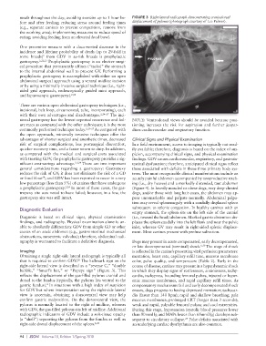
that is required to confirm GDV. The hallmark sign on the color, pulse quality, and temperature (Table 2). Early in the
28
right-side lateral view is described as a “reverse C,” “double course of disease, canines may present in a hyperdynamic shock
bubble,” “Smurf’s hat,” or “Popeye sign” (Figure 3). This in which they display signs of restlessness, anxiousness, tachy-
reflects the displacement of the gas-filled pylorus cranial and cardia, tachypnea, bounding femoral pulses, injected or hyper-
dorsal to the fundus (typically, the pylorus lies ventral to the emic mucous membranes, and rapid capillary refill times. As
gastric fundus). In situations with a high index of suspicion compensatory mechanisms fail and early decompensated shock
4
for GDV but where interpretation using the right-side lateral ensues, dogs progress to having depressed mentation; tachycar-
view is uncertain, obtaining a dorsoventral view may help dia (faster than 140 bpm); rapid and shallow breathing; pale
confirm gastric malposition. On the dorsoventral view, the mucous membranes; prolonged CRT (longer than 3 seconds);
pylorus is normally located to the right of midline, whereas weak and rapid, palpable femoral pulses; and cool extremities.
with GDV, the gas-filled pylorus sits left of midline. Additional During this stage, hypotension (systolic blood pressures lower
radiographic indicators of GDV include a soft-tissue opacity than 90mmHg and MAPs lower than 65mmHg) develops sub-
(a “shelf”) separating the pylorus from the fundus as well as sequent to circulatory collapse. Pulse deficits associated with
29
right-side dorsal displacement of the spleen. 4,28 an underlying cardiac dysrhythmia are also common.
94 | JSOM Volume 18, Edition 1/Spring 2018