
Page 120 - JSOM Winter 2017
P. 120
■ Best the pupil and prevent pupillary block (which can
o Initiate a detailed ocular evaluation and continue lead to angle closure and elevated IOP). 11
monitoring IOP, vision, and RAPD. o Initiate pain control as needed; avoid nonsteroidal
o Continue to check for recurrence of elevated IOP anti-inflammatory drugs, because of risk of worsen-
even after LCC. If the vision deteriorates and the ing intraocular bleeding.
eye again becomes firm after LCC, this may signify o Prevent further injury with antiemetics (ondansetron
rebleeding in the orbit. Evacuation of orbital hem- 4mg ODT/IV/IO/IM every 8 hours as needed).
orrhage is not feasible in a PFC environment and re- o Activate evacuation with goal of evaluation by an
bleeding will require medical treatment. 8 eye surgeon within 24 hours.
• Acetazolamide: 500mg PO initial dose, followed Initiate teleconsultation with photographs.
by 250mg PO 4 times per day (Note: contraindi- ■ Best
cated in patients with sickle cell trait) o Initiate a detailed ocular evaluation to direct treatment.
• If acetazolamide is not available or if the patient Hyphema (anterior chamber injury) 11
cannot take PO, either 3% hypertonic saline o Topical corticosteroid drop (prednisolone acetate
250mL IV or mannitol: 1g/kg IV over 30–60 min- 1%) 4 times per day
utes can be used to decrease IOP. 9 o Cycloplegic eye drop (cyclopentolate 1%), 1 drop
• Corticosteroid: 1g methylprednisolone IV once 10 every 8 hours
Initiate real-time video telemedicine consultation. o Monitor for rebleeding when the clot in anterior
• No altitude restrictions are required for evacuation. chamber retracts, usually at 3–5 days after injury.
NOTES: This may result in vision change and increased size
■ LCC is a vision-saving procedure with minimal risk of of hyphema. 12
causing additional ocular injury. When in doubt, per- o If there is evidence of further bleeding or increasing
form the LCC immediately. IOP, initiate medications to decrease IOP:
■ In thermal burns, consider early LCC (before full OCS • Timolol 0.5%, 1 drop twice a day in affected eye
develops). Fluid resuscitation requirements will take pre- • Acetazolamide 500mg PO initial dose, followed
cedence over the use of medical treatments to reduce IOP. by 250mg PO 4 times per day (Note: contrain-
3. Blunt/closed globe injury. This category includes anterior dicated in patients with sickle cell trait) or 3%
segment injuries such as hyphema (bleeding into the ante- hypertonic saline 250mL IV or mannitol: 1g/kg
rior chamber) and posterior segment injuries such as vitre- IV over 30–60 minutes.
ous hemorrhage and retinal detachment. Blunt trauma can
result in severe loss of vision. NOTE: Tranexamic acid for prevention of rebleeding
in hyphema has not shown any benefit but may be
13
used in multitrauma patients if otherwise indicated.
Posterior chamber injury: Injuries to the retina and
optic nerve as a result of blunt injury will result in vi-
sion loss. Findings may include decreased visual acu-
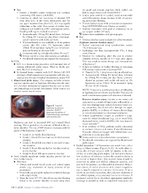
Figure 8 Layered
hyphema in anterior ity, vision loss, loss of red reflex through the pupil,
chamber. (©2017 positive RAPD, or evidence of vitreous hemorrhage
American Academy of or retinal detachment on ultrasound evaluation.
Ophthalmology, reprinted o Initiate supplemental oxygen as available if suspi-
with permission.)
cious for retinal detachment (e.g., cut in visual field,
decreased vision, positive RAPD); this may improve
Hyphema can lead to increased IOP and corneal blood visual outcome. 14
staining. This is graded on the amount of blood in the an- o If no evidence of OGI, perform careful ultrasound
terior chamber. The risk of IOP elevation increases with the to evaluate vitreous and retina, if available/trained.
grade of the hyphema (Figure 8). 11
Transmit ultrasound images with telemedicine con-
• Grade 0: no visible blood layering sultation to an eye specialist.
• Grade 1: blood fills less than one-third of anterior Initiate real-time video telemedicine consultation.
chamber o No altitude restrictions are required for blunt/closed
• Grade 2: blood fills one-third to one-half of ante- globe injury.
rior chamber 4. Eyelid laceration. Lid lacerations can result from either
• Grade 3: blood fills one-half to less than total an- sharp or blunt trauma (Figures 9–11). As with other in-
terior chamber juries, the primary concern with lid injuries is the pos-
• Grade 4: blood fills entire anterior chamber sibility of underlying globe injury. Lid lacerations have a
➤ Goal: Identify significant ocular injuries; protect the eye low incidence of infection (unless the causative factor is
from further injury. an animal or human bite). Any avulsed tissue should be
■ Minimum preserved in saline and chilled, whenever possible, and
o Obtain and record visual acuity and critical injury sent with the patient—not discarded or debrided. Meticu-
details (e.g., mechanism of injury, presence of eye lous closure of eyelid structures with proper magnifica-
protection). tion is usually required to maintain lid function. If fat is
o Protect the injured globe and prevent further damage visible in an eyelid laceration, this indicates violation of
with a rigid shield. the orbital septum, a key anatomic barrier to infection. If
o Raise the head 30°–45°; this allows any free-floating prolapsed orbital fat is identified, appropriate antibiotic
blood in the anterior chamber to settle away from coverage is needed as well as expedited evacuation for
118 | JSOM Volume 17, Edition 4/Winter 2017