
Page 118 - JSOM Winter 2017
P. 118
significant eye injury may be penetrating periocular trauma without full ophthalmic training and equipment. All potential
or lid lacerations, a peaked or teardrop pupil, or abnormal vision-threatening injuries should be evacuated with a goal of
anterior chamber depth. care by an eye surgeon within 24 hours. In some cases, with
prompt teleconsultation or video consultation, it may be safe
Estimation of intraocular pressure (IOP) is essential in inju- to delay evacuation to reduce the effect on the operational sit-
ries such as retrobulbar hemorrhage, but is contraindicated in uation while providing necessary ophthalmic care. Evacuation
injuries with obvious or suspected open globe injury (OGI). within 24 hours is not possible in all situations; therefore, the
If no OGI is suspected, IOP can be estimated using a two- goal of teleconsultation and forward care is to reduce morbid-
finger method. Using the index finger of each hand, gently ity and achieve the best possible outcome. In some operational
apply alternating pressure on the globe through closed lids. environments, optometrists may be available to provide addi-
There should be mild indentation of the eye with normal IOP tional care or consultation closer to the point of injury.
(normal range, 10–21mmHg). With increased IOP, the globe
will be much firmer when compared to the opposite eye, or the Ocular examinations have many specialized components that
examiner’s own eye. The orbit around the eye may also feel a specialist may request. An example template with explana-
tense in a retrobulbar hemorrhage. tion can be found in Appendix C Basic Ocular Examination.
A more detailed examination of the eye can be facilitated by Specific Conditions
the use of a direct ophthalmoscope for magnification. The ex-
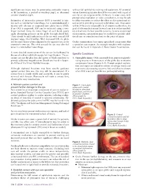
aminer can use the plus lens dial (green or black numbers) to 1. Open globe injury. OGIs can result from penetrating/perfo-
provide additional magnification. Details are found in Appen- rating trauma or from rupture of the globe due to massive
dix B Use of the Direct Ophthalmoscope. compressive forces (Figures 1–5). Prompt surgical explora-
tion and repair are crucial to restore or salvage vision and
Although deployed locations may have specific guidance to prevent a devastating outcome. Safe and effective closure
against contact lens use, this may still be encountered. If a of an OGI is not yet feasible in a prehospital setting.
contact lens is clearly visible and accessible, it can be gently
removed with forceps. Fluorescein will stain a contact lens,
allowing for easy visualization.
4. Maintain patient comfort and Figure 1 Open globe
prevent further damage to the eye. injury with corneal
Pain control is an important component of care in ocular in- laceration, abnormal
juries. Standard Tactical Combat Casualty Care (TCCC) pain pupil shape, and blood
control guidance applies to ocular injuries, including analge- in anterior chamber.
(Photograph by
sic doses of ketamine if needed in systemic polytrauma. Ad- COL Mark Reynolds.)
ditional guidance for pain control in PFC may be found in the
CPG “Analgesia and Sedation Management During Prolonged
Field Care.” 3
Pain is not always present with serious eye injuries, and lack of
pain should not be interpreted as lack of injury. Figure 2 Central
corneal laceration and lid
Ocular injuries can cause a great deal of anxiety for patients, laceration (due to large
and this may affect care. A benzodiazepine may be added to intraocular foreign body).
the treatment plan for anxiety control and facilitation of care (Photograph by
COL Mark Reynolds.)
(diazepam 10 mg by mouth [PO] every 6 hours as needed).
A traumatized eye is highly susceptible to further damage;
antiemetic medications are essential to prevent retching and
increased pressure that can have significant effects on visual
outcome.
Figure 3 Multiple, deep
corneal lacerations, found
Patching both eyes to decrease sympathetic eye movements has to be closed globe injury
not been shown to improve visual outcome. Occluding both on surgical exploration.
eyes will render the patient unable to move independently, (Photograph by
may increase anxiety, and may put the patient and provider COL Mark Reynolds.)
at increased risk in any PFC environment. The use of standard ➤ Goals: Prevent further damage to the eye, prevent infec-
eye protection during patient transport can reduce the risk of tion in the eye (endophthalmitis), and evacuate to an eye
further ocular injury or injury to the fellow eye. Eye protection surgeon as soon as possible.
can be used over a rigid shield to provide increased protection. ■ Minimum
o Maintain high suspicion for OGI; treat any suspected
5. Establish contact with eye care specialist open globe as an open globe until surgical explora-
and prioritize evacuation. tion is available.
Determining the full extent of ophthalmic injuries and the o Obtain and record visual acuity from the injured and
resultant threat of permanent loss of vision is challenging noninjured eye.
116 | JSOM Volume 17, Edition 4/Winter 2017