
Page 62 - Journal of Special Operations Medicine - Spring 2017
P. 62
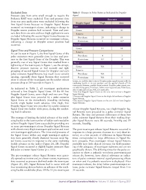
Excluded Data                                      Table 2  Changes in Pulse Status as Indicated by Doppler
          Pressure  data  from  arms  small  enough  to require  the   Signal
          Pediatric RMT were excluded. Time and pressure data                             Extremity
          from one arm application were excluded following the   Doppler Signal    Thigh            Arm
          first Signal Gones  because no Doppler Signal Return
          occurred on tourniquet release, indicating a change in   First
          Doppler sensor position had occurred. Time and pres-    Gones              30             30
          sure data from one arm and two thigh applications were     Returns
          excluded following the second Signal Gones because no      Prerelease      29             19
          Doppler Signal Returns occurred on tourniquet release,
          indicating  a  change  in  Doppler  sensor  position  had      Postrelease  1             10*
          occurred.                                           Second
                                                                Gones                15             14
          Signal Time and Pressure Comparisons                  Returns
          As can be seen in Figure 1, the first Signal Gones of the
          pulse oximeters were generally close in time and pres-     Prerelease      4               3
          sure to the first Signal Gone of the Doppler. This was      Postrelease   10 †             9 ‡
          generally true of any Signal Gones that resulted from a   Third
          tightening of the tourniquet; in Figure 1, see the abrupt,     Gones      NA               1
          discrete pressure increase at 162 seconds and tight
          grouping of second Signal Gones for Doppler and each     Returns
          pulse oximeter. Signal Returns had much more variable      Prerelease     NA               0
          spacing, especially those Signal Returns that occurred      Postrelease   NA               1
          prior to release of the tourniquet; see the ratchet release   Gones, no audible distal arterial Doppler pulse signal present with the
          pressure drop at 209 seconds in Figure 1.          ratcheting buckle in its rest position and the applier’s hands off the
                                                             tourniquet; Returns, audible distal arterial Doppler pulse signal pres-
          As indicated in Table 2, all  tourniquet applications   ent after being gone; Prerelease, before tourniquet release; Postrelease,
                                                             after tourniquet release; NA, not applicable.
          achieved a first Doppler Signal Gone. Of the 60 first   *One first Doppler Signal Gone on the arm did not have a Doppler
          Doppler Signal Gones, seven thigh and two arm Dop-  Signal Return.
                                                              One second Doppler Signal Gone on the thigh did not have a Doppler
          pler Signal Gones were preceded by a pulse oximeter   † Signal Return.
          Signal Gone on the immediately preceding ratcheting   ‡ Two second Doppler Signal Gones on the arm did not have Doppler
          buckle single ladder tooth advance. One thigh, first   Signal Returns.
          Doppler Signal Gone was preceded by a pulse oximeter
          Signal Gone two ladder teeth prior, during the ratchet-  release Doppler Signal Returns, two thigh Doppler Sig-
          ing buckle advance.                                nal Returns were preceded by a pulse oximeter Signal
                                                             Return. The time and pressure differences of those two
          The strategy of limiting the initial advance of the ratch-  pulse oximeter Signal Returns from their trailing Dop-
          eting buckle to the least number of ladder teeth needed to   pler Signal Returns were 88 seconds, 9mmHg and 59
          achieve Doppler Signal Gone succeeded in providing one   seconds, 5mmHg.
          or more pre–tourniquet release Doppler Signal Returns
          with almost every thigh tourniquet application and most   The post–tourniquet release Signal Returns occurred in
          arm tourniquet applications. The times and pressures of   response to a large pressure decrease in a very short in-
          the Signal Gones within any single tourniquet applica-  terval, so a few seconds of difference in Signal Return
          tion were generally closely clustered because most Sig-  times corresponded to large differences in Signal Re-
          nal Gones occurred in response to the same ratcheting   turn pressures. Post–tourniquet release Signal Returns
          buckle advance on the ladder (Figure 2A, 2B). Doppler   probably have little clinical relevance for field monitor-
          Signal Gones occurred at slightly higher pressures than   ing of the adequacy of tourniquet tightness and are not
          did pulse oximeter Signal Gones (p = .011).        graphically shown. The differing times for the pulse ox-
                                                             imeters to show a pulsatile waveform post–tourniquet
          The pre–tourniquet release Signal Returns were gener-  release do, however, have clinical relevance as indica-
          ally spread out in time and, to a lesser extent, in pressure;   tors of pulse oximeter internal signal-processing effects
          they occurred as pressure declined under the tourniquet   on when and if a pulsatile waveform is displayed. After
          (Figure 2C, 2D). Signal Returns had to occur with all   tourniquet release, the delay before post–tourniquet re-
          four monitoring devices before an advance of the ratch-  lease, pulse oximeter Signal Returns ranged from 1 to
          eting buckle would take place. Of the 55 pre–tourniquet   31 seconds.


          40                                      Journal of Special Operations Medicine  Volume 17, Edition 1/Spring 2017