
Page 60 - Journal of Special Operations Medicine - Spring 2017
P. 60
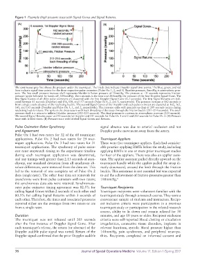
Figure 1  Example thigh pressure trace with Signal Gones and Signal Returns.
            (A)                                                (B)


          The continuous grey line shows the pressure under the tourniquet. The black dots indicate Doppler signal time points. The blue, green, and red
          lines indicate signal time points for the three respective pulse oximeters (Pulse Ox 1, 2, and 3). Baseline pressure, 0mmHg, is atmospheric pres-
          sure. The first small pressure-increase shelf represents bladder inflation pressure of 12mmHg. The pressure at −11 seconds represents friction-
          pressure (spike followed by hands off, 139mmHg). Zero seconds is the time and 285mmHg the pressure of the first Doppler Signal Gone. The
          first Signal Gones of all three pulse oximeters are clustered with the first Doppler Signal Gone (0–3 seconds). The first Signal Returns are indi-
          cated between 11 seconds (Doppler) and 136, 138, and 157 seconds (Pulse Ox 1, 2, and 3, respectively). The pressure increase at 162 seconds is
          from a single tooth advance of the ratcheting buckle. The second Signal Gones of the Doppler and each pulse oximeter are clustered at 162, 163,
          165, and 165 seconds (Doppler and Pulse Ox 3, 1, and 2, respectively). The pressure spike with precipitous drop at 209 seconds occurs during
          ratcheting buckle release. The spike in the drop occurs with back threading of the strap through the friction buckle (211–214 seconds). The small
          pressure shelf is a return to inflated bladder pressure (220–223 seconds). The final pressure is a return to atmospheric pressure (224 seconds).
          The second Signal Returns occur at 210 seconds for Doppler and 211 seconds for Pulse Ox 1 and 2 and 225 seconds for Pulse Ox 3. (A) Pressure
          trace with labeled times. (B) Pressure trace with labeled Signal Gones and Returns.
          Pulse Oximeter–Rater Synchrony                     signal absence was due to arterial occlusion and not
          and Agreement                                      Doppler probe movement away from the artery.
          Pulse Ox 1 had two raters for 32 of the 60 tourniquet
          applications. Pulse Ox 2 had two raters for 29 tour-  Tourniquet Appliers
          niquet applications. Pulse Ox 3 had two raters for 31   There were five tourniquet appliers. Each had consider-
          tourniquet applications. The synchrony of pulse oxim-  able practice applying RMTs before the study, including
          eter rater stopwatch timing to the experiment timeline   applying RMTs in one or more prior tourniquet studies
          during each tourniquet application was determined,   for four of the appliers. There was also an applier assis-
          and any timings with greater than 2.13 seconds of asyn-  tant. The applier assistant pulled directly upward on the
          chrony, one standard deviation from all synchrony ab-  tourniquet handle while the applier pulled the strap di-
          solute differences, were removed from the data set. This   rectly downward, around the limb through the friction
          led to the removal of one complete set of Pulse Ox 2   buckle. This assistance is not essential but was expected
          data (single rater). The other four data set removals for   to aid the achievement of friction-pressures greater than
          asynchrony were from pulse oximeters with two raters;   100mmHg. 6
          the synchronous data sets were retained. Synchronous-
          rater pulse oximeter timing agreement was 82.5% for   Tourniquet Recipients
          calling Signal Gones within 2 seconds of each other and   Tourniquet recipients were volunteers familiar with the
          80.5% for calling Signal Returns within 3 seconds of   tourniquet study through a research course. They were a
          each other. Therefore, the times and associated pressures   convenience sample of students and instructors. Recipi-
          reported either are the averages from two raters or are   ent inclusion criteria were participation in a previous
          from a single rater.                               tourniquet study or participation in the related research
                                                             course, ability to lie down and remain relaxed for 30
          Duration                                           minutes, and age 18 years or older. Recipient exclusion
          The tourniquet  was not released until 205 seconds   criteria were self-reported blood clotting or circulation
          after  the  first  instance  of Doppler  Signal  Gone.  After   irregularities, connective tissue disorders, implants in
          each tourniquet’s release, the return (or absence) of the   relevant locations, systolic blood pressure higher than
          Doppler audible pulse signal was noted. Return of the   140mmHg, pain syndromes, and peripheral neuropa-
          Doppler signal confirmed that the prior Doppler  audible   thies. Recipients completed an informed consent and


          38                                      Journal of Special Operations Medicine  Volume 17, Edition 1/Spring 2017