
Page 78 - JSOM Spring 2026
P. 78
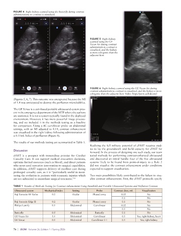
FIGURE 4  Right kidney scanned using the Butterfly during contrast
          administration; no contrast is visualized.


                                                             FIGURE 5  Right kidney
                                                             scanned using the GE
                                                             Vscan Air during contrast
                                                             administration; contrast is
                                                             visualized, and the kidney
                                                             is more echogenic than the
                                                             adjacent liver.


                                                             FIGURE 6  Right kidney scanned using the GE Vscan Air during
                                                             contrast administration; contrast is visualized, and the kidney is more
                                                             echogenic than the adjacent liver. Video: https://jsom.us/kidcont2
          (Figures 5, 6, 7). This outcome was unexpected because the MI
          of 1.4 was anticipated to destroy the perflutren microbubbles.

          The GE Venue is a cart-based portable ultrasound system pres-
          ent in the emergency department of the MTF where the authors
          are stationed. It is not a system typically found in the deployed
          environment. However, it has more powerful image process-
          ing, and we included it in the methods testing as a baseline
          for comparison. Using a 4C curvilinear probe on abdominal
          settings, with an MI adjusted to 0.14, contrast enhancement
          was visualized in the right kidney following administration of
          a 0.11mL bolus of perflutren (Figure 8).
          The results of our methods testing are summarized in Table 1.
                                                             Realizing the full military potential of cFAST requires stud-
                                                             ies to lay the groundwork and build capacity for cFAST far
          Discussion
                                                             forward. In the process of designing one such study, our team
          cFAST is a prospect with tremendous promise for Combat   tested methods for performing contrast-enhanced ultrasound
          Casualty Care. It can support medical evacuation decisions,   and discovered an initial hurdle: four of the five ultrasound
          optimize limited resources (such as blood), and direct patients   systems likely to be found from point-of-injury to a Role 2
          who most need operative intervention to surgical capabilities.   did not visualize the contrast enhancement under conditions
          In addition, cFAST supports delivery of excellent care during   expected to support visualization.
          prolonged casualty care, as it is “particularly useful in moni-
          toring the evolution in patients with traumatic injuries which   Two main possibilities likely contributed to the failure to visu-
          are not submitted to immediate surgical treatment.” 10  alize contrast enhancement. First, the cFAST protocols specify
          TABLE 1  Results of Methods Testing for Contrast-enhancement Using Handheld and Portable Ultrasound Systems and Perflutren Contrast
           Ultrasound system   Mechanical index   Setting        Probe       Contrast dose, mL  Visualization
           Fuji Sonosite M-Turbo    0.2          Ocular        Phased array       0.02             No
                                                                                  0.1              No
           Fuji Sonosite Edge II    0.2          Ocular        Phased array       0.1              No
           Philips Lumify           0.1         Abdominal      Curvilinear        0.02             No
                                                                                  0.1              No
           Butterfly                0.5         Abdominal       Butterfly         0.1              No
           GE Vscan Air             1.4         Abdominal      Curvilinear        0.1       Yes; right kidney, heart
           GE Venue                 0.1         Abdominal     Curvilinear 5-1     0.1         Yes; right kidney

          76  |  JSOM   Volume 26, Edition 1 / Spring 2026