
Page 82 - JSOM Spring 2025
P. 82
Model Preparation for Cricothyrotomy and the DVM and MD groups performing CTT, the DVM and MD
Tube Tracheostomy groups performing TT, and in each group comparing their
Evaluators prepared cadavers and study areas and laid out sur- performance of CTT and TT. Five experimenters per group
gical instruments for each technique to ensure standardization performed each procedure and were rated/scored by two inde-
between both groups. Cadavers were positioned and secured in pendent observers.
dorsal recumbency. A rolled towel was placed under the dorsal
aspect of the neck to mildly hyperextend the cranial cervical Results
region; this facilitated laryngeal cartilage exposure and tra-
cheal palpation by pushing the larynx and trachea to a more Ten fresh frozen, ethically sourced cadavers weighing 55–106
superficial position. The ventral aspect of the neck was clipped pounds (24.9–48.1kg) were used for the study (Table 1). The ex-
extending caudally from the ramus of the mandible to the tho- perimenters consisted of five emergency medicine physician resi-
racic inlet and approximately 3–4 inches (7.62–10.16cm) on dents (area of concentration, AOC 62A) and five commissioned
each side of the midline. A brief surgical site preparation with VCOs (area of concentration, AOC 64A). All experimenters
70% isopropyl alcohol was performed over the clipped area to enrolled in the study completed the study. Medical residents
remove clipped hair and any gross debris from the surgical site. (MD group) had on average two years of experience practicing
For the second technique, if a large incision was made during medicine. In comparison, only one of the veterinarians (DVM
the first technique, the skin was pulled across the midline and group) had five years of experience and the other DVM exper-
secured with towel clamps so that underlying anatomy was not imenters were newly commissioned recent graduates of a pro-
visible for the second technique. fessional Doctor of Veterinary Medicine degree program. Each
experimenter performed a CTT and TT in a crossover fashion
Recording and Scoring resulting in 20 procedures performed on 10 cadavers.
Procedural timing for CTT and TT commenced from the first
incision and ceased when the participant verbally indicated Time to Complete the Procedure
completion with the word “stop.” Technique success was The DVM group experimenters completed CTT at different
defined by confirming the tip of the CTT or TT tube seated times. The longest time to complete was 622 seconds and the
within the airway. Complications were determined and re- shortest time to complete was 20 seconds, which was by the
corded by examination from the investigators. Damage scores VCO with five years of experience. The MD group completed
were provided for each injury identified; scores were adapted CTT faster than the DVM group. There was minimal variabil-
from previously conducted veterinary studies. Upon comple- ity in the time to complete CTT between the experimenters in
19
tion of the first round of techniques by a participant, only a the MD and DVM groups; however, due to the high standard
partial dissection of the cadaver by the investigators was per- deviation (SD) within the DVM group, the time difference to
formed to confirm tube placement and any gross luminal or complete CTT between the two groups was not statistically
laryngeal damage. Following the completion of the second significant (DVM: 239.6 [SD 251.661] s vs. MD: 37.4 [SD
round of techniques, the investigators opened the entire cervi- 11.283] s; Figure 1A). Although the DVM group completed TT
cal airway on the midline for a complete examination of gross slightly slower than the MD group, the time difference was not
damage to the airway structures. statistically significant (DVM: 133.4 [SD 88.002] s vs. MD: 91
[SD 41.635] s). The time to complete CTT for the MD group
To evaluate the technical difficulty of CTT and TT, each par- was statistically shorter than the time to complete TT (CTT:
ticipant completed a standardized post-study questionnaire 37.4 [SD 11.283] s vs. TT: 91 [SD 41.635] s, P<.05). Despite
that was adopted from prior similar studies. 19,20 The question- the overall faster achievement of TT by the DVM group, there
naire consisted of: was no statistically significant difference between the time to
complete CTT and TT (CTT: 239.6 [SD 251.661] s vs. TT:
1. A visual analogue scale (VAS), scored from 0 to 10 with 133.4 [SD 88.002] s).
0 being “the easiest” and 10 being “the most technically
challenging.” Post-procedure Damage Score
2. Questions indicating which technique the participant Two independent observers reviewed each procedure (CTT or
would select in practice if faced with a “cannot intubate, TT) performed by the MD and DVM groups using a dam-
cannot oxygenate” (CICO) event in an MWD. age scoring system extrapolated from Hardjo et al. as shown
16
in Box 4. The MD group did not cause gross damage to the
Statistical Analyses trachea, surrounding muscles, or esophagus while performing
All data were analyzed with GraphPad Prism 10.2.1 ( GraphPad CTT. Except for the only experienced veterinarian, the remain-
Software, Boston, MA). Alpha for statistical significance was ing DVM experimenters caused some damage while perform-
set at 0.05. Numerical values are reported as mean and SD. ing CTT. The difference between the mean damage score of the
Unpaired t test was employed to compare differences between DVM and the MD groups was statistically significant (DVM:
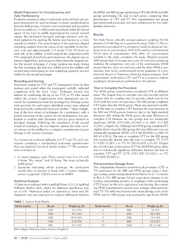
TABLE 1 Cadaver Body Weights
Model number Breed Weight, kg (lb) Model number Breed Weight, kg (lb)
1 GSD 35.4 (78) 6 BM 25 (55)
2 GSD 38.1 (84) 7 GSD 40.4 (89)
3 GSD 45.4 (100) 8 GSD 31.4 (69)
4 GSD 31.8 (70) 9 BM 30.5 (67)
5 BM 26.4 (58) 10 GSD 41.2 (106)
BM = Belgian Malinois; GSD = German Shepherd.
80 | JSOM Volume 25, Edition 1 / Spring 2025