
Page 81 - JSOM Spring 2025
P. 81
proficiency for human patients, emergency human healthcare instruments, and materials were standardized between both
clinicians have limited time in their schedule to learn a novel, groups and both techniques. Each experimenter was randomly
technical, and perishable skill such as the TT. In particular assigned to a cadaver pair to perform both techniques on each
since their use of TT would be limited to MWDs with UAO. cadaver. Experimenters performed their first attempt of each
Having the ability to readily translate the training, skills, and technique on a fresh cadaver while their second attempt was
knowledge they already possess for performing CTT in people on the same paired cadavers, but the techniques were crossed
to MWDs prevents the need and time associated with learning over.
the TT technique. Furthermore, training U.S. Army Veterinary
Corps Officers (VCOs) in a technique that is the least tech- Description of Techniques
nical, affords the greatest skills retention and is the most ex- Techniques for CTT and TT were standardized for the experi-
pedient for achieving emergent airway access. This approach menters. For CTT, experimenters demonstrated the rapid four-
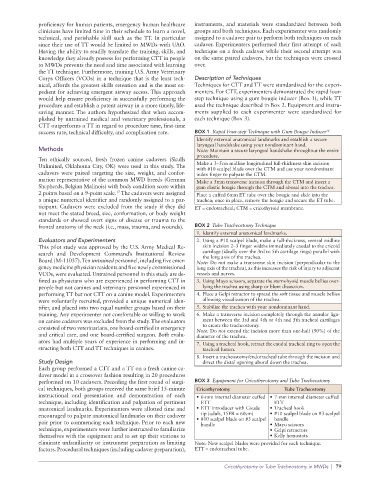
would help ensure proficiency in successfully performing the step technique using a gum bougie inducer (Box 1), while TT
procedure and establish a patent airway in a more timely, life- used the technique described in Box 2. Equipment and instru-
saving manner. The authors hypothesized that when accom- ments supplied to each experimenter were standardized for
plished by untrained medical and veterinary professionals, a each technique (Box 3).
CTT outperforms a TT in regard to procedure time, first-time
success rate, technical difficulty, and complication rate. BOX 1 Rapid Four-step Technique with Gum Bougie Inducer 18
Identify external anatomical landmarks and establish a secure
laryngeal handshake using your nondominant hand.
Methods Note: Maintain a secure laryngeal handshake throughout the entire
procedure.
Ten ethically sourced, fresh frozen canine cadavers (Skulls
Unlimited, Oklahoma City, OK) were used in this study. The Make a 3–5cm midline longitudinal full-thickness skin incision
cadavers were paired targeting the size, weight, and confor- with #10 scalpel blade over the CTM and use your nondominant
index finger to palpate the CTM.
mation representative of the common MWD breeds (German Make a 5mm transverse incision through the CTM and insert a
Shepherds, Belgian Malinois) with body condition score within gum elastic bougie through the CTM and aboral into the trachea.
2 points based on a 9-point scale. The cadavers were assigned Place a cuffed 6mm ET tube over the bougie and slide into the
17
a unique numerical identifier and randomly assigned to a par- trachea; once in place, remove the bougie and secure the ET tube.
ticipant. Cadavers were excluded from the study if they did ET = endotracheal; CTM = cricothyroid membrane.
not meet the stated breed, size, conformation, or body weight
standards or showed overt signs of disease or trauma to the
frontal anatomy of the neck (i.e., mass, trauma, and wounds). BOX 2 Tube Tracheostomy Technique
1. Identify external anatomical landmarks.
Evaluators and Experimenters 2. Using a #10 scalpel blade, make a full-thickness, ventral midline
This pilot study was approved by the U.S. Army Medical Re- skin incision 2–3 finger widths immediately caudal to the cricoid
search and Development Command’s Institutional Review cartilage (ideally over the 3rd to 5th cartilage rings) parallel with
the long axis of the trachea.
Board (M-11037). Ten untrained personnel, including five emer- Note: Do not make a transverse skin incision (perpendicular to the
gency medicine physician residents and five newly commissioned long axis of the trachea), as this increases the risk of injury to adjacent
VCOs, were evaluated. Untrained personnel in this study are de- vessels and nerves.
fined as physicians who are experienced in performing CTT in 3. Using Mayo scissors, separate the sternohyoid muscle bellies over-
people but not canines and veterinary personnel experienced in lying the trachea using sharp or blunt dissection.
performing TT but not CTT on a canine model. Experimenters 4. Place a Gelpi retractor to spread the soft tissue and muscle bellies
were voluntarily recruited, provided a unique numerical iden- allowing visualization of the trachea.
tifier, and placed into two equal number groups based on their 5. Stabilize the trachea with your nondominant hand.
training. Any experimenter not comfortable or willing to work 6. Make a transverse incision completely through the annular liga-
on canine cadavers was excluded from the study. The evaluators ment between the 3rd and 4th or 4th and 5th tracheal cartilages
consisted of two veterinarians, one board-certified in emergency to create the tracheostomy.
and critical care, and one board-certified surgeon. Both evalu- Note: Do not extend the incision more than one-half (50%) of the
diameter of the trachea.
ators had multiple years of experience in performing and in- 7. Using a tracheal hook, retract the caudal tracheal ring to open the
structing both CTT and TT techniques in canines. tracheal lumen.
8. Insert a tracheostomy/endotracheal tube through the incision and
Study Design direct the distal opening aboral down the trachea.
Each group performed a CTT and a TT on a fresh canine ca-
daver model in a crossover fashion resulting in 20 procedures
performed on 10 cadavers. Preceding the first round of surgi- BOX 3 Equipment for Cricothyrotomy and Tube Tracheostomy
cal techniques, both groups received the same brief 15- minute Cricothyrotomy Tube Tracheostomy
instructional oral presentation and demonstration of each • 6-mm internal diameter cuffed • 7-mm internal diameter cuffed
technique, including identification and palpation of pertinent ETT ETT
anatomical landmarks. Experimenters were allotted time and • ETT introducer with Coude • Tracheal hook
encouraged to palpate anatomical landmarks on their cadaver tip (adult, 15FR × 60cm) • #10 scalpel blade on #3 scalpel
handle
pair prior to commencing each technique. Prior to each new • #10 scalpel blade on #3 scalpel • Mayo scissors
handle
technique, experimenters were further instructed to familiarize • Gelpi retractors
themselves with the equipment and to set up their stations to • Kelly hemostats
eliminate unfamiliarity or instrument preparation as limiting Note: New scalpel blades were provided for each technique.
factors. Procedural techniques (including cadaver preparation), ETT = endotracheal tube.
Cricothyrotomy or Tube Tracheostomy in MWDs | 79