
Page 97 - JSOM Winter 2022
P. 97
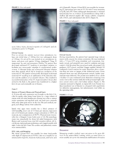
FIGURE 3  Chest radiograph.                        of 6.12mmol/L. Repeat 12-lead ECG was notable for worsen-
                                                                 ing ST depressions now seen in V3–V6 with T-wave inversion
                                                                 in II, III, and aVF. Chest radiograph demonstrated a widened
                                                                 mediastinum but no evidence of pneumothorax along with a
                                                                 shallow left internal jugular (IJ) venous catheter, orogastric
                                                                 tube (OGT), and endotracheal tube (ETT) (Figure 4).
                                                                 FIGURE 4  Chest radiograph.


              acute kidney injury, elevated troponin of 2.65ng/mL and ele-
              vated lactic acid of 3.79mg/dL.
              Clinical Course
              Prior to arrival, the patient received three  amiodarone bo-  Clinical Course
              luses (an initial dose of 300mg then two subsequent doses   During resuscitation, the patient had repeated large volume
              of 150mg). On arrival he was started on an amiodarone in-  emesis with concern for airway protection. He was intubated
              fusion and given oral aspirin 325mg, clopidogrel 75mg, IV   with a 7.5-mm ETT using etomidate and rocuronium. Fen-
              magnesium 2g, and oral potassium 20mEq. The patient then   tanyl, versed, and propofol were used for sedation and pain
              developed sustained VT resulting in procedural sedation to   control. A left IJ central line was placed under ultrasound (US)
              facilitate  two  unsuccessful  attempts  in  synchronized  cardio-  guidance, OGT placed, and Bair Hugger applied. Bedside US
              version. A lidocaine bolus and subsequent lidocaine infusion   confirmed clinical concern for aortic dissection after a dissec-
              were then started, which led to temporary resolution of the   tion flap was seen on images of the abdominal aorta. Given
              sustained VT. The patient subsequently developed recalcitrant   adequate heart rate and blood pressure control, further inter-
              sustained VT resulting in an uptitration of the lidocaine infu-  ventions were deferred. The patient was medevac’d to a Role
              sion and the addition of an esmolol infusion. This resulted in   3 facility. CT scan confirmed aortic dissection extending from
              successful termination of his VT. Approximately 6 hours after   aortic root to iliac arteries with clot in the proximal right com-
              arrival to the Role 2, the patient was transferred to a higher   mon carotid artery and a left kidney infarct (Figure 5). His
              level of care and eventually out of theater.       troponin increased to 8ng/mL on repeat lab draw. Patient was
                                                                 then transferred out of theater to a hospital with capabilities
                                                                 for definitive management, but unfortunately died while on
              Case 3
                                                                 the operating table.
              History of Present Illness and Physical Exam
              A 58-year-old male contractor was brought to the Role 2 fa-  FIGURE 5  CT scan.
              cility by medics after being found down in a large volume of
              emesis with left-sided hemiparesis. Patient was able to answer
              yes/no questions and reported neck and back pain currently
              with some chest pain earlier in the day. His past medical, sur-
              gical, and allergy history were unknown.

              Initial vital signs were notable for a blood pressure of
              126/52mmHg, heart rate of 53 beats per minute, respiratory
              rate of 16 breaths per minute, oxygen saturation of 100% on
              room air, and a temperature of 34.9°C. Physical exam was no-
              table for a Glasgow Coma Scale score of 14. His neurologic
              exam was remarkable for dense left-sided hemiparesis with
              flexion and increased tone, total left sensation deficit, left fa-
              cial droop, rightward gaze preference, and sluggish pupils with
              right 2 mm and left 4 mm. His cardiac and pulmonary exams
              were unremarkable.

                                                                 Discussion
              ECG, Labs, and Imaging
              His initial 12-lead ECG was notable for sinus bradycardia   Managing complex medical cases can prove to be quite dif-
              with slight ST depressions in V5/V6. Labs were notable for an   ficult in the austere Role 2 setting, which, as noted above, is
              undetectable troponin, moderate leukocytosis, and lactic acid   most suitably set up for the immediate stabilization of trauma

                                                                          Complex Medical Patients in Role 2 Environment  |  95