
Page 18 - JSOM Winter 2022
P. 18
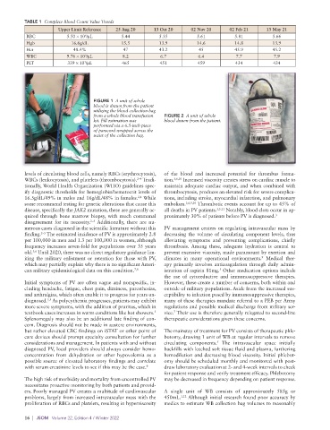
TABLE 1  Complete Blood Count Value Trends
                      Upper Limit Reference  25 Aug 20   13 Oct 20     02 Nov 20     02 Feb 21    13 May 21
           RBC           5.50 × 10 /μL       5.44          5.35          5.61          5.81          5.66
                                6
           Hgb             16.6g/dL          15.5          13.9          14.6          14.8          13.9
           Hct              48.4%             47           43.2           45           45.9          45.2
           WBC           9.76 × 10 /μL        8.2           6.7          6.4           7.7           7.9
                                3
           PLT            339 × 10 /μL        465          451           459           434           434
                                3


                                   FIGURE 1  A unit of whole
                                   blood is drawn from the patient
                                   utilizing the blood collection bag
                                   from a whole blood transfusion   FIGURE 2  A unit of whole
                                   kit. Fill estimation was   blood drawn from the patient.
                                   performed via a 6.5-inch piece
                                   of paracord wrapped across the
                                   waist of the collection bag.


          levels of circulating blood cells, namely RBCs (erythrocytosis),   of the blood and increased potential for thrombus forma-
          WBCs (leukocytosis), and platelets (thrombocytosis).  Tradi-  tion. 3,6,10  Increased viscosity creates stress on cardiac muscle to
                                                   2–4
          tionally, World Health Organization (WHO) guidelines spec-  maintain adequate cardiac output, and when combined with
          ify diagnostic thresholds for hemoglobin/hematocrit levels of   thrombocytosis, produces an elevated risk for severe complica-
                                                   2,4
          16.5g/dL/49% in males and 16g/dL/48% in females.  While   tions, including stroke, myocardial infarction, and pulmonary
          some recommend testing for genetic alterations that cause this   embolism. 3,6,9,10  Thrombotic events account for up to 45% of
          disease, specifically the JAK2 mutation, these are generally ac-  all deaths in PV patients. 3,5,11  Notably, blood clots occur in ap-
          quired through bone marrow biopsy, with much communal   proximately 30% of patients before PV is diagnosed. 3
          disagreement for its necessity.   Additionally, there are nu-
                                  2–5
          merous cases diagnosed in the scientific literature without this   PV management centers on regulating intravascular mass by
                2–5
          finding.  The estimated incidence of PV is approximately 2.8   decreasing the volume of circulating component levels, thus
          per 100,000 in men and 1.3 per 100,000 in women, although   alleviating symptoms and preventing  complications,  chiefly
          frequency increases seven-fold for populations over 35 years   thrombosis.  Among these, adequate hydration is central to
          old.  Until 2020, there was no direct regulatory guidance lim-  prevent excessive viscosity, made paramount by exertion and
             3,6
                                                                                                 9
          iting the military enlistment or retention for those with PV,   climates in many operational environments.  Medical ther-
          which may partially explain why there is no significant Ameri-  apy primarily involves anticoagulation through daily admin-
          can military epidemiological data on this condition. 7,8  istration of aspirin 81mg.  Other medication options include
                                                                                 5
                                                             the use of cytoreductive and immunosuppressive therapies.
          Initial symptoms of PV are often vague and nonspecific, in-  However, these create a number of concerns, both within and
          cluding headache, fatigue, chest pain, dizziness, paresthesias,   outside of military populations. Aside from the increased sus-
          and arthralgias, which often enable it to progress for years un-  ceptibility to infection posed by immunosuppressive therapies,
          diagnosed.  As polycythemia progresses, patients may exhibit   many of these therapies mandate referral to a PEB per Army
                  3,5
          more severe symptoms, with the addition of pruritus, which in   regulations and possible medical discharge from military ser-
                                                                7
                                                         5
          textbook cases increases in warm conditions like hot showers.    vice.  Their use is therefore generally relegated to second-line
          Splenomegaly may also be an additional late finding of con-  therapeutic considerations given these concerns.
          cern. Diagnosis should not be made in austere environments,
          but rather elevated CBC findings on iSTAT or other point of   The mainstay of treatment for PV consists of therapeutic phle-
          care devices should prompt specialty consultation for further   botomy, drawing 1 unit of WB at regular intervals to remove
          considerations and management. In patients with and without   circulating components.   The intravascular space initially
                                                                                5
          diagnosed PV, local providers should always consider hemo-  backfills with leeched soft tissue fluid and plasma, furthering
          concentration from dehydration or other hypovolemia as a   hemodilution and decreasing blood viscosity. Initial phlebot-
          possible source of elevated laboratory findings and correlate   omy should be scheduled monthly and monitored with post-
          with serum creatinine levels to see if this may be the case. 9  draw laboratory evaluation at 2- and 4-week intervals to check
                                                             for patient response and verify treatment efficacy. Phlebotomy
          The high risk of morbidity and mortality from uncontrolled PV   may be decreased in frequency depending on patient response.
          necessitates proactive monitoring by both patients and provid-
          ers. Poorly managed PV creates a multitude of cardiovascular   A single unit of  WB consists of approximately 585g or
          problems, largely from increased intravascular mass with the   450mL. 1,12  Although initial research found poor accuracy by
          proliferation of RBCs and platelets, resulting in hyperviscosity   medics to estimate WB collection bag volumes to reasonably

          16  |  JSOM   Volume 22, Edition 4 / Winter 2022