
Page 58 - JSOM Summer 2022
P. 58
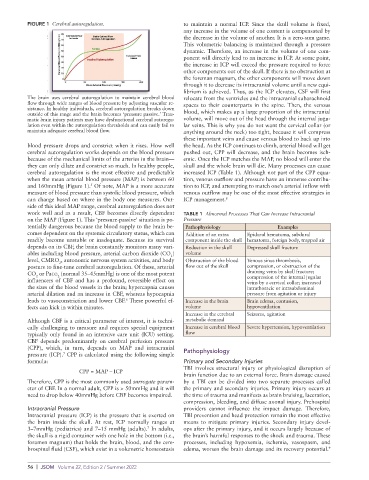
FIGURE 1  Cerebral autoregulation.                 to  maintain  a  normal  ICP.  Since  the  skull  volume  is  fixed,
                                                             any increase in the volume of one content is compensated by
                                                             the decrease in the volume of another. It is a zero-sum game.
                                                             This volumetric balancing is maintained through a pressure
                                                             dynamic. Therefore, an increase in the volume of one com-
                                                             ponent will directly lead to an increase in ICP. At some point,
                                                             the increase in ICP will exceed the pressure required to force
                                                             other components out of the skull. If there is no obstruction at
                                                             the foreman magnum, the other components will move down
                                                             through it to decrease its intracranial volume until a new equi-
                                                             librium is achieved. Thus, as the ICP elevates, CSF will first
          The brain uses cerebral autoregulation to maintain cerebral blood   relocate from the ventricles and the intracranial subarachnoid
          flow through wide ranges of blood pressure by adjusting vascular re-  spaces  to  their counterparts  in the  spine.  Then,  the venous
          sistance. In healthy individuals, cerebral autoregulation breaks down
          outside of this range and the brain becomes ‘pressure passive.’ Trau-  blood, which makes up a large proportion of the intracranial
          matic brain injury patients may have dysfunctional cerebral autoregu-  volume, will move out of the head through the internal jugu-
          lation even within the autoregulation thresholds and can easily fail to   lar veins. This is why you do not want the cervical collar (or
          maintain adequate cerebral blood flow.             anything around the neck) too tight, because it will compress
                                                             these important veins and cause venous blood to back up into
          blood pressure drops and constrict when it rises. How well   the head. As the ICP continues to climb, arterial blood will get
          cerebral autoregulation works depends on the blood pressure   pushed out, CPP will decrease, and the brain becomes isch-
          because of the mechanical limits of the arteries in the brain—  emic. Once the ICP matches the MAP, no blood will enter the
          they can only dilate and constrict so much. In healthy people,   skull and the whole brain will die. Many processes can cause
          cerebral autoregulation is the most effective and predictable   increased ICP (Table 1). Although not part of the CPP equa-
          when the mean arterial blood pressure (MAP) is between 60   tion, venous outflow and pressure have an immense contribu-
          and 160mmHg (Figure 1).  Of note, MAP is a more accurate   tion to ICP, and attempting to match one’s arterial inflow with
                               5
          measure of blood pressure than systolic blood pressure, which   venous outflow may be one of the most effective strategies in
          can change based on where in the body one measures. Out-  ICP management. 8
          side of this ideal MAP range, cerebral autoregulation does not
          work well and as a result, CBF becomes directly dependent   TABLE 1  Abnormal Processes That Can Increase Intracranial
          on the MAP (Figure 1). This ‘pressure-passive’ situation is po-  Pressure
          tentially dangerous because the blood supply to the brain be-  Pathophysiology    Examples
          comes dependent on the systemic circulatory status, which can   Addition of an extra   Epidural hematoma, subdural
          readily become unstable or inadequate. Because its survival   component inside the skull  hematoma, foreign body, trapped air
          depends on its CBF, the brain constantly monitors many vari-  Reduction in the skull   Depressed skull fracture
          ables including blood pressure, arterial carbon dioxide (CO )   volume
                                                         2
          level, CMRO , autonomic nervous system activities, and body   Obstruction of the blood   Venous sinus thrombosis,
                    2
          posture to fine-tune cerebral autoregulation. Of these, arterial   flow out of the skull  compression, or obstruction of the
          CO  or Paco  (normal 35–45mmHg) is one of the most potent               draining veins by skull fracture;
             2
                    2
          influencers of CBF and has a profound, reversible effect on             compression of the internal jugular
                                                                                  veins by a cervical collar; increased
          the sizes of the blood vessels in the brain; hypercapnia causes         intrathoracic or intraabdominal
          arterial dilation and an increase in CBF, whereas hypocapnia            pressure from agitation or injury
          leads to vasoconstriction and lower CBF.  These powerful ef-  Increase in the brain   Brain edema, contusion,
                                          6
          fects can kick in within minutes.                   volume              hypoventilation
                                                              Increase in the cerebral   Seizures, agitation
          Although CBF is a critical parameter of interest, it is techni-  metabolic demand
          cally challenging to measure and requires special equipment   Increase in cerebral blood   Severe hypertension, hypoventilation
          typically only found in an intensive care unit (ICU) setting.   flow
          CBF depends predominantly on cerebral perfusion pressure
          (CPP), which, in turn, depends on MAP and intracranial
                      7
          pressure (ICP).  CPP is calculated using the following simple   Pathophysiology
          formula:                                           Primary and Secondary Injuries
                                                             TBI involves structural injury or physiological disruption of
                           CPP = MAP – ICP
                                                             brain function due to an external force. Brain damage caused
          Therefore, CPP is the most commonly used surrogate param-  by a TBI can be divided into two separate processes called
          eter of CBF. In a normal adult, CPP is > 50mmHg and it will   the primary and secondary injuries. Primary injury occurs at
          need to drop below 40mmHg before CBF becomes impaired.  the time of trauma and manifests as brain bruising, laceration,
                                                             compression, bleeding, and diffuse axonal injury. Prehospital
          Intracranial Pressure                              providers cannot influence the impact damage. Therefore,
          Intracranial pressure (ICP) is the pressure that is exerted on   TBI prevention and head protection remain the most effective
          the brain inside the skull. At rest, ICP normally ranges at   means  to mitigate  primary  injuries.  Secondary  injury devel-
                                                 7
          3–7mmHg (pediatrics) and 7–15 mmHg (adults).  In adults,   ops after the primary injury, and it occurs largely because of
          the skull is a rigid container with one hole in the bottom (i.e.,   the brain’s harmful responses to the shock and trauma. These
          foramen magnum) that holds the brain, blood, and the cere-  processes, including hypoxemia, ischemia, vasospasm, and
                                                                                                            9
          brospinal fluid (CSF), which exist in a volumetric homeostasis   edema, worsen the brain damage and its recovery potential.

          56  |  JSOM   Volume 22, Edition 2 / Summer 2022