
Page 59 - JSOM Summer 2022
P. 59
Fortunately, secondary injuries are potentially preventable and example, if you tell someone the patient has a GCS of 7, that
treatable. Therefore, mitigation of secondary injuries and pre- person will quickly understand that this patient is sick even
vention of potentiators of those injuries have been the focus of without knowing the entire neurological exam.
the modern TBI management.
There is not one physical examination method that fits all TBI
TBI patients often cannot produce a reliable history. In such patients. Medics and prehospital care providers must tailor the
cases, medical providers need to have a high index of suspi- exam based on the patient’s clinical stability, injury type(s), degree
cion for conditions that may have caused the accident (e.g., of cooperation from the patient, use of sedatives and paralytics,
stroke, seizure, hypoglycemia, etc.). Remember that it takes a and the operational environment and constraints. Nevertheless,
tremendous amount of energy to fracture the skull. In fact, a the following is a neuro-focused examination that a provider
skull fracture increases the probability of a surgical intracra- should consider at a minimum when managing a TBI patient:
nial injury by 20-fold in a comatose patient and a 400-fold in
10
a conscious patient. Approximately 25–60% of TBI patients Visual Inspection
involved in major accidents also have significant concurrent • Evidence of a basal skull fracture, which indicates high
11
spinal injuries, with tendency for C1–C3 involvement. Care- impact injury with potential cranial nerve and vascu-
less handling of these patients can cause devastating injuries lar injuries: periorbital ecchymosis (raccoon’s eyes sign),
and even death. Therefore, always assume that TBI patients postauricular ecchymosis (Battle’s sign), CSF rhinorrhea/
have a spinal injury until proven otherwise. In addition, unre- otorrhea, hemotympanum, or laceration of the ear canal
sponsive patients have a 50–65% probability of having inju- • Evidence of facial fractures, which is commonly associated
ries to one or more organ systems, so treat the whole patient with TBI and can compromise neurological exam, causes
and not just the TBI. 12 disorientation and agitation in patients, causes CSF leak
through separation of the face from the skull, and creates
airway issues: assess for Le Forte fractures by palpating the
Diagnostics
facial bones including the zygomatic arch and the orbital rim
Examination • Physical signs of spinal trauma: look for bruising and
TBI is a clinical diagnosis. Therefore, physical examination and deformity
neurological assessment are absolutely critical. Serial neurolog- • Intermittent or continuous clinical seizures
ical exam is useful for monitoring disease progression, treat- • Cranial nerves
ment monitoring, and prognosis in the setting of TBI. Despite o Optic nerve: In a conscious patient, ask the patient to
many limitations, GCS score remains the most widely accepted read any printed material. If unable, ask to count fin-
and validated system for objectively assessing the level of con- gers. If unable, check light perception. If unconscious,
sciousness in TBI patients (Table 2). Generally, GCS scores of check for afferent pupillary defect by conducting the
14–15 are considered mild TBI, 9–13 are moderate, and ≤ 8 are swinging flashlight test.
severe TBI. This stratification is arbitrary and oversimplified, o Pupil: Check for size in ambient light and test reaction
with each category containing highly variable clinical pictures. to light on both sides.
Nevertheless, it is a simple system that helps with standardiza- o Abducens nerve: Palsy will present as a medially devi-
13
tion. The GCS is very useful in trending the progression of the ated eye. Abducens palsy may indicate skull base frac-
patient (getting better, worse, or staying the same), and letting ture or increased ICP.
other providers know how severe or well your patient is. For o Facial nerve: Check for facial symmetry. Skull base frac-
ture can lead to facial nerve injury.
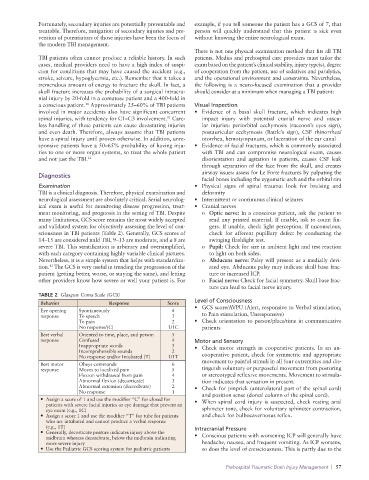
TABLE 2 Glasgow Coma Scale (GCS)
Behavior Response Score Level of Consciousness
Eye opening Spontaneously 4 • GCS score/AVPU (Alert, responsive to Verbal stimulation,
response To speech 3 to Pain stimulation, Unresponsive)
To pain 2 • Check orientation to person/place/time in communicative
No response/(C) 1/1C patients
Best verbal Oriented to time, place, and person 5
response Confused 4 Motor and Sensory
Inappropriate words 3 • Check motor strength in cooperative patients. In an un-
Incomprehensible sounds 2
No response and/or Intubated (T) 1/1T cooperative patient, check for symmetric and appropriate
Best motor Obeys commands 6 movement to painful stimuli in all four extremities and dis-
response Moves to localized pain 5 tinguish voluntary or purposeful movement from posturing
Flexion withdrawal from pain 4 or stereotyped reflexive movements. Movement to stimula-
Abnormal flexion (decorticate) 3 tion indicates that sensation in present.
Abnormal extension (decerebrate) 2 • Check for pinprick (anterolateral part of the spinal cord)
No response 1 and position sense (dorsal column of the spinal cord).
• Assign a score of 1 and use the modifier “C” for closed for • When spinal cord injury is suspected, check resting anal
patients with severe facial injuries or eye damage that prevent an
eye exam (e.g., 1C) sphincter tone, check for voluntary sphincter contraction,
• Assign a score 1 and use the modifier “T” for tube for patients and check for bulbocavernosus reflex.
who are intubated and cannot produce a verbal response
(e.g., 1T) Intracranial Pressure
• Generally, decorticate posture indicates injury above the
midbrain whereas decerebrate, below the midbrain indicating • Conscious patients with worsening ICP will generally have
more severe injury headache, nausea, and frequent vomiting. As ICP worsens,
• Use the Pediatric GCS scoring system for pediatric patients so does the level of consciousness. This is partly due to the
Prehospital Traumatic Brain Injury Management | 57