
Page 60 - JSOM Summer 2022
P. 60
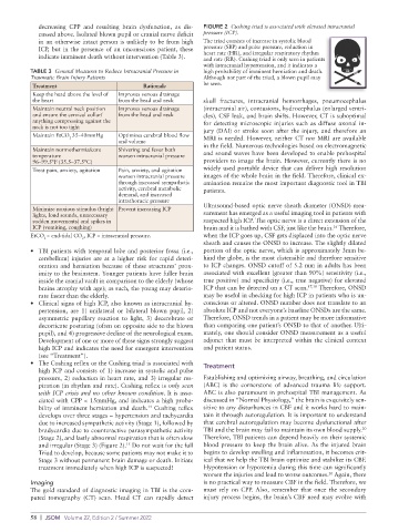
decreasing CPP and resulting brain dysfunction, as dis-  FIGURE 2  Cushing triad is associated with elevated intracranial
            cussed above. Isolated blown pupil or cranial nerve deficit   pressure (ICP).
            in an otherwise intact person is unlikely to be from high   The triad consists of increase in systolic blood
            ICP, but in the presence of an unconscious patient, these   pressure (SBP) and pulse pressure, reduction in
            indicate imminent death without intervention (Table 3).  heart rate (HR), and irregular respiratory rhythm
                                                             and rate (RR). Cushing triad is only seen in patients
                                                             with intracranial hypertension, and it indicates a
          TABLE 3  General Measures to Reduce Intracranial Pressure in   high probability of imminent herniation and death.
          Traumatic Brain Injury Patients                    Although not part of the triad, a blown pupil may
           Treatment                       Rationale         be seen.
           Keep the head above the level of   Improves venous drainage
           the heart                from the head and neck   skull  fractures,  intracranial  hemorrhages,  pneumocephalus
           Maintain neutral neck position   Improves venous drainage   (intracranial air), contusions, hydrocephalus (enlarged ventri-
           and ensure the cervical collar/  from the head and neck  cles), CSF leak, and brain shifts. However, CT is suboptimal
           anything compressing against the                  for detecting microscopic injuries such as diffuse axonal in-
           neck is not too tight                             jury (DAI) or stroke soon after the injury, and therefore an
           Maintain EtCO 35–40mmHg  Optimizes cerebral blood flow   MRI is needed. However, neither CT nor MRI are available
                     2
                                    and volume
           Maintain normothermia/core   Shivering and fever both   in the field. Numerous technologies based on electromagnetic
           temperature              worsen intracranial pressure  and sound waves have been developed to enable prehospital
           96–99.5°F (35.5–37.5°C)                           providers to image the brain. However, currently there is no
           Treat pain, anxiety, agitation  Pain, anxiety, and agitation   widely used portable device that can deliver high resolution
                                    worsen intracranial pressure   images of the whole brain in the field. Therefore, clinical ex-
                                    through increased sympathetic   amination remains the most important diagnostic tool in TBI
                                    activity, cerebral metabolic   patients.
                                    demand, and increased
                                    intrathoracic pressure
           Minimize noxious stimulus (bright  Prevent increasing ICP  Ultrasound-based optic nerve sheath diameter (ONSD) mea-
           lights, loud sounds, unnecessary                  surement has emerged as a useful imaging tool in patients with
           sudden movements) and spikes in                   suspected high ICP. The optic nerve is a direct extension of the
                                                                                                   16
           ICP (vomiting, coughing)                          brain and it is bathed with CSF, just like the brain.  Therefore,
          EtCO = end-tidal CO , ICP = intracranial pressure.  when the ICP goes up, CSF gets displaced into the optic nerve
              2          2
                                                             sheath and causes the ONSD to increase. The slightly dilated
          •  TBI patients with temporal lobe and posterior fossa (i.e.,   portion of the optic nerve, which is approximately 3mm be-
            cerebellum) injuries are at a higher risk for rapid deteri-  hind the globe, is the most distensible and therefore sensitive
            oration and herniation because of these structures’ prox-  to ICP changes. ONSD cutoff of 5.2 mm in adults has been
            imity to the brainstem. Younger patients have fuller brain   associated with excellent (greater than 90%) sensitivity (i.e.,
            inside the cranial vault in comparison to the elderly (whose   true positive) and specificity (i.e., true negative) for elevated
            brains atrophy with age); as such, the young may deterio-  ICP that can be detected on a CT scan. 17,18  Therefore, ONSD
            rate faster than the elderly.                    may be useful in checking for high ICP in patients who is un-
          •  Clinical signs of high ICP, also known as intracranial hy-  conscious or altered. ONSD number does not translate to an
            pertension,  are  1) unilateral  or bilateral  blown  pupil, 2)   absolute ICP and not everyone’s baseline ONSDs are the same.
            asymmetric pupillary reaction to light, 3) decerebrate or   Therefore, ONSD trends in a patient may be more informative
            decorticate posturing (often on opposite side to the blown   than comparing one patient’s ONSD to that of another. Ulti-
            pupil), and 4) progressive decline of the neurological exam.   mately, one should consider ONSD measurement as a useful
            Development of one or more of these signs strongly suggest   adjunct  that  must  be  interpreted  within  the  clinical  context
            high ICP and indicates the need for emergent intervention   and patient status.
            (see “Treatment”).
          •  The Cushing reflex or the Cushing triad is associated with   Treatment
            high ICP and consists of 1) increase in systolic and pulse
            pressure, 2) reduction in heart rate, and 3) irregular res-  Establishing and optimizing airway, breathing, and circulation
            piration (in rhythm and rate). Cushing reflex is only seen   (ABC) is the cornerstone of advanced trauma life support.
            with ICP crisis and no other known condition. It is asso-  ABC is also paramount in prehospital TBI management. As
            ciated with CPP < 15mmHg, and indicates a high proba-  discussed in “Normal Physiology,” the brain is exquisitely sen-
            bility of imminent herniation and death.  Cushing reflex   sitive to any disturbances in CBF and it works hard to main-
                                             14
            develops over three stages – hypertension and tachycardia   tain it through autoregulation. It is important to understand
            due to increased sympathetic activity (Stage 1), followed by   that cerebral autoregulation may become dysfunctional after
                                                                                                            20
            bradycardia due to counteractive parasympathetic activity   TBI and the brain may fail to maintain its own blood supply.
            (Stage 2), and lastly abnormal respiration that is often slow   Therefore, TBI patients can depend heavily on their systemic
                                      15
            and irregular (Stage 3) (Figure 2).  Do not wait for the full   blood pressure to keep the brain alive. As the injured brain
            Triad to develop, because some patients may not make it to   begins to develop swelling and inflammation, it becomes crit-
            Stage 3 without permanent brain damage or death. Initiate   ical that we help the TBI brain optimize and stabilize its CBF.
            treatment immediately when high ICP is suspected!  Hypotension or hypoxemia during this time can significantly
                                                             worsen the injuries and lead to worse outcomes.  Again, there
                                                                                                  20
          Imaging                                            is no practical way to measure CBF in the field. Therefore, we
          The gold standard of diagnostic imaging in TBI is the com-  must rely on CPP. Also, remember that once the secondary
          puted tomography (CT) scan. Head CT can rapidly detect   injury process begins, the brain’s CBF need may evolve with


          58  |  JSOM   Volume 22, Edition 2 / Summer 2022