
Page 80 - JSOM Winter 2021
P. 80
We maintained general anesthesia with 2–2.5% isoflurane We delivered pulmonary contusions as described above. Subse-
in 40–100% oxygen to maintain an oxygen saturation of quently, we subjected swine to tibial fracture using a benchtop
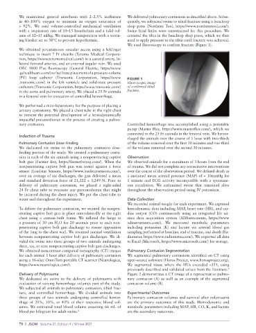
> 92%. We used volume-controlled mechanical ventilation shop press (Northern Tool, https://www.northerntool.com/).
with a respiratory rate of 10–15 breaths/min and a tidal vol- Swine hind limbs were unrestrained for this procedure. We
ume of 12–15 ml/kg. We managed temperature with a warm- centered the tibia in the benchtop shop press, which we then
ing blanket set to 38°C to prevent hypothermia. used to apply pressure to the tibia until fracture was achieved.
We used fluoroscopy to confirm fracture (Figure 1).
We obtained percutaneous vascular access using a Seldinger
technique to insert 7 Fr sheaths (Terumo Medical Corpora-
tion, https://www.terumomedical.com/) in a carotid artery, bi-
lateral femoral arteries, and an external jugular vein. We used
OEC 9800 Plus fluoroscopy (General Electric, https://www
.gehealthcare.com/) to facilitate placement of a pressure- volume
(PV) loop catheter (Transonic Corporation, https://www FIGURE 1
.transonic.com/) in the left ventricle and solid-state pressure Fluoroscopic image
catheters (Transonic Corporation, https://www.transonic.com/) of confirmed tibial
in the aorta and pulmonary artery. We placed a 25 Fr cannula fracture.
in a femoral vein for execution of controlled hemorrhage.
We performed a mini-laparotomy for the purpose of placing a
urinary cystostomy. We placed a chest tube in the right chest
to prevent the potential development of a hemodynamically
impactful pneumothorax in the process of creating a pulmo-
nary contusion. Controlled hemorrhage was accomplished using a peristaltic
pump (Master Flex, https://www.masterflex.com/), which we
connected to the 25 Fr cannula in the femoral vein. We hemor-
Induction of Trauma
rhaged the animals over the course of 1 hour with two-thirds
Pulmonary Contusion Dose-Finding of the volume removed over the first 30 minutes and one-third
We dedicated six swine to the pulmonary contusion dose- of the volume removed over the second 30 minutes.
finding portion of the study. We created a pulmonary contu-
sion in each of the six animals using a nonpenetrating captive Observation
bolt gun (Farmer Boy, https://farmerboyag.com/). When the We observed animals for a maximum of 3 hours from the end
nonpenetrating captive bolt gun was tested against a force of trauma. We did not complete any resuscitative interventions
sensor (Loadstar Sensors, https://www.loadstarsensors.com/), over the course of the observation period. We defined death as
over an average of six discharges, the gun delivered a mean a sustained mean arterial pressure (MAP) of < 10mmHg for
and standard deviation force of 21,322 ± 3,249 N. Prior to 1 minute and ECG activity incompatible with a spontane-
delivery of pulmonary contusion, we placed a right-sided ous circulation. We euthanized swine that remained alive
24 Fr chest tube to evacuate any pneumothorax that might throughout the observation period using IV potassium.
be incurred during the chest injury. We put the chest tube to
water seal throughout the experiment. Data Collection
We recorded animal weight for each experiment. We captured
To deliver the pulmonary contusion, we secured the nonpen- hemodynamic data including MAP, heart rate (HR), and car-
etrating captive bolt gun in place anterolaterally at the right diac output (CO) continuously using an integrated life sci-
chest using a custom-built frame. We inflated the lungs to ence data acquisition system (ADInstruments, https://www
a pressure of 30 cm H O for 20 seconds prior to each non- adinstruments.com/). We measured metabolic parameters
2
penetrating captive bolt gun discharge to ensure apposition including potassium (K) and lactate via arterial blood gas
of the lung to the chest wall. We resumed normal ventilation sampling performed at baseline, end of trauma, and death (Ra-
between nonpenetrating captive bolt gun discharges. We di- diometer, https://www.radiometer.com/). We exported all data
vided the swine into three groups of two animals undergoing to Excel (Microsoft, https://www.microsoft.com/) for storage.
three, six, or nine nonpenetrating captive bolt gun discharges.
We obtained noncontrast computed tomography (CT) images Pulmonary Contusion Segmentation
for each animal 1 hour after delivery of pulmonary contusion We segmented pulmonary contusions identified on CT using
using a 16-slice OmniTom portable CT scanner (Neurologica, open-source software (Horos Project, www.horosproject.org).
https://www.neurologica.com/). We segmented tissue where the HUs exceeded –351, using
previously described and validated values from the literature.
9
Delivery of Polytrauma Figure 2 demonstrates a CT image of a representative pulmo-
We dedicated six swine to the delivery of polytrauma with nary contusion (A) as well as an example of the segmented
evaluation of varying hemorrhage volumes part of the study. contusion volume (B).
We subjected all animals to pulmonary contusion, tibial frac-
ture, and controlled hemorrhage. We divided animals into Experimental Outcomes
three groups of two animals undergoing controlled hemor- Pulmonary contusion volumes and survival after polytrauma
rhage of 20%, 30%, or 40% of their respective blood vol- are the primary outcomes of this study. Hemodynamic and
umes. We estimated total blood volume assuming 66 mL of metabolic parameters including MAP, HR, CO, K, and lactate
blood per kilogram for adult swine. 8 are the secondary outcomes.
78 | JSOM Volume 21, Edition 4 / Winter 2021