
Page 92 - JSOM Spring 2021
P. 92
approved by the FDA. We selected a pulse rate of 2 to 5 for was transferred to a local hospital per current medical rules of
our patients, using lower pulse rates for extremity imaging and engagement. Because of the radiologic evidence available, we
higher pulse rates for axial imaging. This provides a radia- felt comfortable observing the patient without further surgical
tion dose of 0.052 to 0.18 mSv, which is comparable to previ- interventions. This allowed us to remain ready for subsequent
ously reported radiation dosages for standard imaging studies casualties, preserving operating room time and nursing care.
(0.001 to 1.2 mSv). 6
Case No. 2
All images were reviewed and interpreted by a board- certified A 24-year-old man presented for evaluation after having an
emergency medicine physician, board-certified general sur- up-armored door closed on his right index finger while on a
geon, or board-certified orthopedic surgeon. Images were pre- forward-staged mission. The patient is right-hand dominant
viewed immediately after exposure and, if deemed inadequate, and a member of a special operations team. Clinical examina-
were repeated. Given the positioning of our patients, a pro- tion demonstrated a nail bed injury with a partially avulsed
vider often had to hold the generator to provide an adequate nail plate. He was holding his finger in a partially flexed posi-
image and therefore was not the recommended 10 feet away tion at the distal interphalangeal joint. The finger was tempo-
during operations. rarily splinted by the co-located surgical team, and the patient
returned to his forward operating base for further evaluation
and consultation with the FRST orthopedic surgeon. By x-ray,
Clinical Case Scenarios
the surgeon was able to fully evaluate the comminuted, in-
Case No. 1 tra-articular fracture with 100% displacement and apex dorsal
A 28-year-old man presented to the FRST with a gunshot angulation (Figure 3A). After digital block, a closed reduc-
wound to his left, lateral arm. Trauma assessment revealed tion was performed, and the patient’s finger was placed in an
decreased breath sounds to the left chest, and an eFAST (ex- AlumaFoam splint (Hartmann USA). Confirmation of accept-
tended focused assessment with sonography for trauma) ex- able reduction is shown in Figure 3B. At 6 weeks, follow-up
amination was notable for lack of lung sliding in the left chest. radiographs showed appropriate healing (Figure 3C). At this
A 36Fr thoracostomy tube was placed for a suspected hemo- time, the patient was transitioned from a full-time splint to
pneumothorax and returned approximately 400mL of blood. a night-only splint. With field radiographic capabilities, our
While chest tube output was monitored, a secondary survey team was able to treat this soldier without the need for evacu-
demonstrated a suspected proximal humerus fracture. Using ation to the Role 3. Had this resource not been available, the
the radiography capabilities of the EOD team, we were able patient would have required evacuation, adversely affecting
to identify a retained bullet fragment in the left chest and con- his team’s mission and operational capabilities.
firm acceptable placement of the chest tube. We were also able
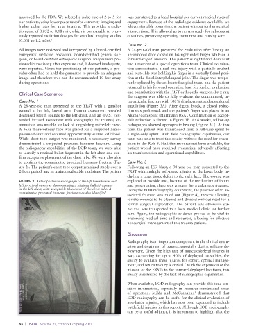
to confirm the comminuted proximal humerus fracture (Fig- Case No. 3
ure 2). The patient’s chest tube output remained stable over a Following an IED blast, a 30-year-old man presented to the
2-hour period, and he maintained stable vital signs. The patient FRST with multiple soft-tissue injuries to the lower body, in-
cluding a large tissue defect to the right heel. The wound was
FIGURE 2 Anteroposterior radiograph of the left hemithorax and explored at bedside and, because of the mechanism of injury
left proximal humerus demonstrating a retained bullet fragment and presentation, there was concern for a calcaneus fracture.
in the left chest, with acceptable placement of the chest tube. A Using the EOD radiography equipment, the presence of an as-
comminuted proximal humerus fracture was also identified.
sociated fracture was ruled out (Figure 4), thereby allowing
for the wounds to be cleaned and dressed without need for a
formal surgical exploration. The patient was otherwise sta-
ble and was transported to a local medical clinic for further
care. Again, the radiographic evidence proved to be vital to
preserving medical time and resources, allowing for effective
nonsurgical management of this trauma patient.
Discussion
Radiography is an important component in the clinical evalu-
ation and treatment of trauma, especially during military de-
ployment. Given the high rate of musculoskeletal injuries in
war, accounting for up to 40% of deployed causalities, the
ability to evaluate these injuries for extent, optimal manage-
ment, and return to duty is critical. With the expansion of the
7
mission of the FRSTs to the forward-deployed locations, this
ability is restricted by the lack of radiographic capabilities.
When available, EOD radiography can provide this time-sen-
sitive information, especially in resource-constrained areas
of operation. Milda and McGranahan demonstrated that
5
EOD radiography can be useful for the clinical evaluation of
non-battle injuries, which has now been expanded to include
battlefield injuries in this report. Although EOD radiography
can be a useful adjunct, it is important to highlight that the
88 | JSOM Volume 21, Edition 1 / Spring 2021