
Page 93 - JSOM Spring 2021
P. 93
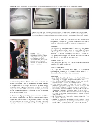
FIGURE 3  Lateral radiographs of the right index finger demonstrating a comminuted, intra-articular fracture of the distal phalanx.

                                    (A)


                                            (B)                                                           (C)


                                          (A) Distal phalanx with 100% fracture displacement and apex dorsal angulation. (B) Post-reduction
                                          lateral radiograph demonstrating the comminuted, intra-articular fracture in acceptable alignment in
                                          an AlumaFoam splint. (C) Six-week follow-up: lateral radiograph demonstrating appropriate healing.

                                                                 being aware of other available resources and proper usage
                                                                 can augment the clinical care provided, ensuring that medical
                                                                 readiness and mission capability are never compromised.

                                                                 Disclaimer
                                                                 The opinions or assertions contained herein are the private
                                                                 views of the authors and are not to be construed as official or
                                                                 reflecting the views of the Department of Defense or US Gov-
                                       FIGURE 4  Clinical image   ernment. The authors are employees of the US Government.
                                       taken while obtaining a lateral   This work was prepared as part of their official duties and, as
                                       radiograph of the right foot   such there is no copyright to be transferred.
                                       for evaluation of possible
                                       calcaneal fracture. Radiographs
                                       demonstrated no fracture,   Financial Disclosure
                                       allowing for expedited patient   The authors have indicated they have no financial relationship
                                       care and evacuation.      related to this article to disclose.
                                                                 Author Contributions
                                                                 CH, SV, RL, and KAS treated the patients. CH, SV, and KAS
                                                                 developed the manuscript. RL provided critical edits. All au-
                                                                 thors read and approved the final manuscript.

                                                                 References
                                                                 1.  Joint Chiefs of Staff. Joint Publication 4-02: Joint Health Services.
                                                                   Washington, DC: National Defense University Press; 2017.
              radiation safety of these devices is not rated for human use.   2.  Cota SA. Refining SOF surgical support to meet joint force de-
                                                                   mand. Combat & Casualty Care. 2018;Spring:8–19.
              Future studies should seek to evaluate the radiation exposure   3.  United States Government US Army. Field Manual 4-02.25. Em-
              of these devices, as well as the implications of image settings   ployment  of  Forward  Surgical  Teams:  Tactics,  Techniques,  and
              on patient dose exposure. Permanent inclusion of portable   Procedures. Washington, DC: Department of the Army; 2003.
              x-ray to the modified table of organization and equipment will   4.  United States Government US Army. Field Manual 4-30. Ordnance
              require further investigation to determine the cost and feasibil-  Operations. Washington, DC: Department of the Army; 2004.
              ity to equip all FSTs/FRSTs.                       5.  Milda GS, McGranahan ME. Explosive ordinance disposal team
                                                                   equipment and its use in diagnosing extremity trauma. Mil Med.
                                                                   2002;167(12):1044.
              In the forward-deployed setting, providers in the FST/FRST   6.  Lin EC. Radiation risk from medical imaging. Mayo Clin Proc.
              are likely to be presented with clinical scenarios in which plain   2010;85(12):1142–1146.
              radiography may be beneficial in optimizing patient care and   7.  Belmont PJ, Owens BD, Schoenfeld AJ. Musculoskeletal injuries
              minimizing excessive resource utilization. Although plain radi-  in Iraq and Afghanistan: epidemiology and outcomes following a
              ography is not a component of the team’s deployed equipment,   decade of war. J Am Acad Orthop Surg. 2016;24(6):341–348.


                                                                        EOD Radiography in the Forward-Deployed Setting  |  89