
Page 33 - JSOM Spring 2021
P. 33
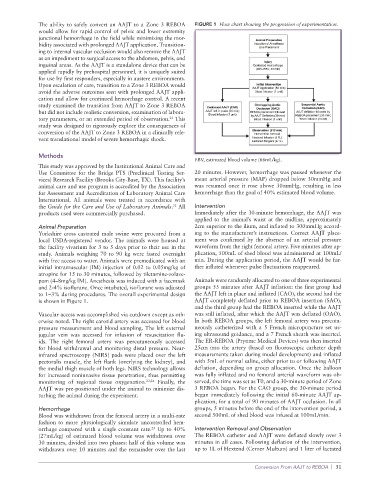
The ability to safely convert an AAJT to a Zone 3 REBOA   FIGURE 1  Flow chart showing the progression of experimentation.
              would allow for rapid control of pelvic and lower extremity
              junctional hemorrhage in the field while minimizing the mor-
              bidity associated with prolonged AAJT application. Transition-
              ing to internal vascular occlusion would also remove the AAJT
              as an impediment to surgical access to the abdomen, pelvis, and
              inguinal areas. As the AAJT is a standalone device that can be
              applied rapidly by prehospital personnel, it is uniquely suited
              for use by first responders, especially in austere environments.
              Upon escalation of care, transition to a Zone 3 REBOA would
              avoid the adverse outcomes seen with prolonged AAJT appli-
              cation and allow for continued hemorrhage control. A recent
              study examined the transition from AAJT to Zone 3 REBOA
              but did not include realistic conversion, examination of labora-
              tory parameters, or an extended period of observation.  This
                                                        21
              study was designed to rigorously explore the consequences of
              conversion of the AAJT to Zone 3 REBOA in a clinically rele-
              vant translational model of severe hemorrhagic shock.

              Methods
                                                                 EBV, estimated blood volume (66mL/kg).
              This study was approved by the Institutional Animal Care and
              Use Committee for the Bridge PTS (Preclinical Testing Ser-  20 minutes. However, hemorrhage was paused whenever the
              vices) Research Facility (Brooks City-Base, TX). This facility’s   mean arterial pressure (MAP) dropped below 30mmHg and
              animal care and use program is accredited by the Association   was resumed once it rose above 30mmHg, resulting in less
              for Assessment and Accreditation of Laboratory Animal Care   hemorrhage than the goal of 40% estimated blood volume.
              International. All animals were treated in accordance with
              the Guide for the Care and Use of Laboratory Animals.  All   Intervention
                                                          22
              products used were commercially purchased.         Immediately after the 30-minute hemorrhage, the AAJT was
                                                                 applied to the animal’s waist at the midline, approximately
              Animal Preparation                                 2cm superior to the ilium, and inflated to 300mmHg accord-
              Yorkshire cross castrated male swine were procured from a   ing to the manufacturer’s instructions. Correct AAJT place-
              local USDA-registered vendor. The animals were housed at   ment was confirmed by the absence of an arterial pressure
              the facility vivarium for 3 to 5 days prior to their use in the   waveform from the right femoral artery. Five minutes after ap-
              study. Animals weighing 70 to 90 kg were fasted overnight   plication, 500mL of shed blood was administered at 100mL/
              with free access to water. Animals were premedicated with an   min. During the application period, the AAJT would be fur-
              initial intramuscular (IM) injection of 0.02 to 0.05mg/kg of   ther inflated whenever pulse fluctuations reappeared.
              atropine for 15 to 30 minutes, followed by tiletamine-zolaze-
              pam (4–8mg/kg IM). Anesthesia was induced with a facemask   Animals were randomly allocated to one of three experimental
              and 2-4% isoflurane. Once intubated, isoflurane was adjusted   groups 55 minutes after AAJT inflation: the first group had
              to 1–3% during procedures. The overall experimental design   the AAJT left in place and inflated (CAO), the second had the
              is shown in Figure 1.                              AAJT completely deflated prior to REBOA insertion (SAO),
                                                                 and the third group had the REBOA inserted while the AAJT
              Vascular access was accomplished via cutdown except as oth-  was still inflated, after which the AAJT was deflated (OAO).
              erwise noted. The right carotid artery was accessed for blood   In both REBOA groups, the left femoral artery was percuta-
              pressure measurement and blood sampling. The left external   neously catheterized with a 5 French micropuncture set us-
              jugular vein was accessed for infusion  of resuscitation  flu-  ing ultrasound guidance, and a 7 French sheath was inserted.
              ids. The right femoral artery was percutaneously accessed   The ER-REBOA (Prytime Medical Devices) was then inserted
              for blood withdrawal and monitoring distal pressure. Near-   25cm into the artery (based on fluoroscopic catheter depth
              infrared spectroscopy (NIRS) pads were placed over the left   measurements taken during model development) and inflated
              pectoralis muscle, the left flank (overlying the kidney), and   with 5mL of normal saline, either prior to or following AAJT
              the medial thigh muscle of both legs. NIRS technology allows   deflation, depending on group allocation. Once the balloon
              for increased noninvasive tissue penetration, thus permitting   was fully inflated and no femoral arterial waveform was ob-
              monitoring of regional tissue oxygenation. 23,24  Finally, the   served, the time was set as T0, and a 30-minute period of Zone
              AAJT was pre-positioned under the animal to minimize dis-  3 REBOA began. For the CAO group, the 30-minute period
              turbing the animal during the experiment.          began immediately following the initial 60-minute AAJT ap-
                                                                 plication, for a total of 90 minutes of AAJT occlusion. In all
              Hemorrhage                                         groups, 5 minutes before the end of the intervention period, a
              Blood was withdrawn from the femoral artery in a multi-rate   second 500mL of shed blood was infused at 100mL/min.
              fashion to more physiologically simulate uncontrolled hem-
              orrhage compared with a single constant rate.  Up to 40%   Intervention Removal and Observation
                                                   25
              (27mL/kg) of estimated blood volume was withdrawn over   The REBOA catheter and AAJT were deflated slowly over 3
              30 minutes, divided into two phases: half of this volume was   minutes in all cases. Following deflation of the intervention,
              withdrawn over 10 minutes and the remainder over the last   up to 1L of Hextend (Cerner Multum) and 1 liter of lactated

                                                                                    Conversion From AAJT to REBOA  |  31