
Page 73 - JSOM Winter 2018
P. 73
model could be used for training to acquire the skills needed
to adequately place an endovascular sheath in a femoral ar- BOX 1
tery model and subsequently place a REBOA catheter in aortic
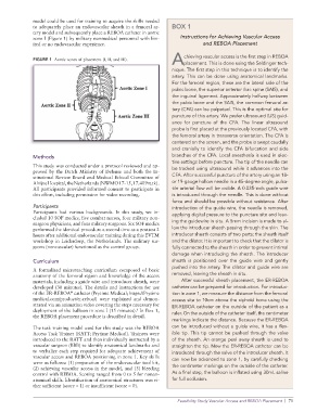
zone I (Figure 1) by military nonmedical personnel with lim- Instructions for Achieving Vascular Access
ited or no endovascular experience. and REBOA Placement
chieving vascular access is the first step in REBOA
FIGURE 1 Aortic zones of placement (I, II, and III).
Aplacement. This is done using the Seldinger tech-
nique. The first step in this technique is to identify the
artery. This can be done using anatomical landmarks.
For the femoral region, these are the lateral side of the
pubic bone, the superior anterior iliac spine (SAIS), and
the inguinal ligament. Approximately halfway between
the pubic bone and the SAIS, the common femoral ar-
tery (CFA) can be palpated. This is the optimal site for
puncture of this artery. We prefer ultrasound (US) guid-
ance for puncture of the CFA. The linear ultrasound
probe is first placed at the previously located CFA, with
the femoral artery in transverse orientation. The CFA is
centered on the screen, and the probe is swept caudally
and cranially to identify the CFA bifurcation and side
Methods branches of the CFA. Local anesthesia is used in elec-
tive settings before puncture. The tip of the needle can
This study was conducted under a protocol reviewed and ap-
proved by the Dutch Ministry of Defense and both the In- be tracked using ultrasound while it advances into the
stitutional Review Board and Medical Ethical Committee of CFA. After successful puncture of the artery using an 18-
Alrijne Hospital, the Netherlands (NWMO 17-15, 17.409rt.tk). or 19-gauge hollow needle in a 45-degree angle, pulsa-
All participants provided informed consent to participate in tile arterial flow will be visible. A 0.035-inch guide wire
this effort, including permission for video recording. is introduced through the needle. This is done without
force and should be possible without resistance. After
Participants introduction of the guide wire, the needle is removed,
Participants had various backgrounds. In this study, we in- applying digital pressure to the puncture site and leav-
cluded 10 SOF medics, five combat nurses, four military non-
surgeon physicians, and four military surgeons. Six SOF medics ing the guidewire in situ. A 5mm incision is made to al-
performed the identical procedure a second time as a posttest 2 low the introducer sheath passing through the skin. The
hours after additional endovascular training during this EVTM introducer sheath consists of two parts: the sheath itself
workshop in Leiderdorp, the Netherlands. The military sur- and the dilator. It is important to check that the dilator is
geons (nonvascular) functioned as the control group. fully connected to the sheath in order to prevent intimal
damage when introducing the sheath. The introducer
Curriculum sheath is positioned over the guide wire and gently
pushed into the artery. The dilator and guide wire are
A formalized microteaching curriculum composed of basic
anatomy of the femoral region and knowledge of the access removed, leaving the sheath in situ.
materials, including a guide wire and introducer sheath, were After successful sheath placement, the ER-REBOA
developed (30 minutes). The details and instructions for use catheter can be prepared for introduction. For introduc-
of the ER-REBOA catheter (Prytime Medical; https://Prytime tion in zone 1, we measure the distance from the femoral
®
medical.com/product/er-reboa/) were explained and demon- access site to 10cm above the xiphoid bone using the
strated via an animation video covering the steps necessary for ER- REBOA catheter on the outside of the patient as a
deployment of the balloon in zone I (15 minutes). In Box 1,
6
the REBOA placement procedure is described in detail. ruler. On the outside of the catheter itself, the centimeter
markings indicate the distance. Because the ER- REBOA
The task training model used for this study was the REBOA can be introduced without a guide wire, it has a flex-
Access Task Trainer (RATT; Prytime Medical). Trainees were ible tip. This tip cannot be pushed through the valve
introduced to the RATT and then individually instructed by a of the sheath. An orange peel away sheath is used to
vascular surgeon (BBB) to identify anatomical landmarks and straighten the tip. Now the ER- REBOA catheter can be
to verbalize each step required for adequate achievement of introduced through the valve of the introducer sheath. It
vascular access and REBOA positioning in zone 1. Key skills can now be advanced to zone 1, by carefully checking
were as follows: (1) preparation of the endovascular tool kit,
(2) achieving vascular access in the model, and (3) bleeding the centimeter markings on the outside of the catheter.
control with REBOA. Scoring ranged from 0 to 5 for nonan- As a final step, the balloon is inflated using 30mL saline
atomical skills. Identification of anatomical structures was ei- for full occlusion.
ther sufficient (score = 1) or insufficient (score = 0).
Feasibility Study Vascular Access and REBOA Placement | 71