
Page 68 - JSOM Summer 2018
P. 68
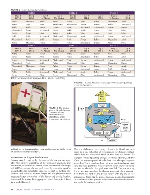
FIGURE 2  Order of surgical procedures.
                        WEEK 1                           WEEK 2                           WEEK 3
             Day 1      Day 2      Day 3      Day 1      Day 2      Day 3      Day 1      Day 2      Day 3
             EPF-1      LCS-1     No Motion  No Motion   LCS-1      EPF-1      EPF-1     No Motion   LCS-1
           Pelvis     Abdominal  Ankle      Ankle      Femur      Pelvis     Pelvis     Abdominal  Ankle
           Femur      Pelvis     Pelvis     Abdominal  Abdominal  Femur      Femur      Ankle      Abdominal
           Ankle      Ankle      Femur      Femur      Pelvis     Abdominal  Abdominal  Femur      Femur
           Abdominal  Femur      Abdominal  Pelvis     Ankle      Ankle      Ankle      Pelvis     Pelvis
           Pelvis     Femur      Pelvis     Ankle      Ankle      Pelvis     Ankle      Pelvis     Pelvis
           Ankle      Ankle      Abdominal  Abdominal  Abdominal  Ankle      Abdominal  Femur      Abdominal
           Abdominal  Pelvis     Ankle      Femur      Pelvis     Femur      Pelvis     Abdominal  Ankle
           Femur      Abdominal  Femur      Pelvis     Femur      Abdominal  Femur      Ankle      Pelvis
                        WEEK 4                           WEEK 5                           WEEK 6
             Day 1      Day 2      Day 3      Day 1      Day 2      Day 3      Day 1      Day 2      Day 3
             EPF-1      LCS-1     No Motion  No Motion   LCS-1      EPF-1      EPF-1     No Motion   LCS-1
           Ankle      Femur      Femur      Pelvis     Ankle      Ankle      Femur      Pelvis     Pelvis
           Pelvis     Ankle      Pelvis     Femur      Femur      Abdominal  Pelvis     Ankle      Ankle
           Femur      ABdominal  Abdominal  Abdominal  Pelvis     Femur      Abdominal  Femur      Abdominal
           Abdominal  Pelvis     Ankle      Ankle      Abdominal  Pelvis     Ankle      Abdominal  Femur
           Abdominal  Abdominal  Femur      Ankle      Femur      Femur      Femur      Pelvis     Ankle
           Femur      Femur      Ankle      Femur      Ankle      Abdominal  Ankle      Ankle      Femur
           Ankle      Ankle      Abdominal  Abdominal  Pelvis     Ankle      Pelvis     Femur      Abdominal
           Pelvis     Pelvis     Pelvis     Pelvis     Abdominal  Pelvis     Abdominal  Abdominal  Pelvis
          EPF, Expeditionary Fast Transport; LCS, Littoral Combat Ship.
                                                             FIGURE 4  Medical Mission Module Support Container operating
                                                             room configuration.


                                        FIGURE 3  The Medical
                                        Mission Module Support
                                        Container aboard
                                        a Stewart motion
                                        table (Moog Series
                                        6DOF500E, Model 170).


          subtask or for team members to sit and recuperate in the event   For the abdominal procedure, reduction in blood loss was
          of a motion sickness incident.                     used as a key indicator of performance for damage control.
                                                             Blood loss was measured based on the flow rate before the
          Assessment of Surgical Performance                 surgeon initiated packing sponges into the abdomen, and this
          Success was determined by the state of the patient surrogate   flow rate was compared with the flow rate when packing was
          after the surgery, regardless of the workload involved, time   complete. The prepacking flow rate was based on elapsed time
          consumed, or number of minor errors committed. The surgi-  and the flow quantities measured from when the blood pump
          cal SMEs evaluated the surgical teams by assessing specific,   started to when the first pack was installed. The postpacking
          quantifiable, and repeatable variables for each of the four pro-  flow rate was based on the elapsed time and blood quantity
          cedures. For instance, incision length and pin placement were   lost from the start of the bowel repair until the end of the
          measured after completion of the femur and pelvic fixation.   procedure. Based on the prepacking and postpacking results,
          Measured data were then aggregated into a five-point Likert-  a calculation of the percent reduction in flow was determined
          type scale (Figure 5).                             using the following equation:


          66  |  JSOM   Volume 18, Edition 2/Summer 2018