
Page 108 - JSOM Summer 2018
P. 108
pathogenic microorganisms. Repetitive self-inflicted trauma Initially, the exudate is a clear (serous) discharge; over time,
1,4
(i.e., licking, chewing, scratching) damages the skin barrier it becomes suppurative and may dry and crust on the surface.
and erodes the superficial dermal surface, creating a favorable Often, the hair appears matted (i.e., glued together from the
environment for bacterial colonization for commensal and exudate) around and over the open wound. Because this is not
5
noncommensal microbial organisms. Over time, bacterial colo- folliculitis or furunculosis, lesions are not usually surrounded
nization causes a secondary bacterial infection. The secondary by pustules; however, small papules (satellite lesions) may be
1
bacterial infection, which is intensely pruritic, further perpetu- present surrounding the lesion. They appear primarily around
2,5
ates the cycle of itch, scratch, and self-mutilation. the lateral aspect of the head, periaural region (i.e., around the
ears), caudal dorsal trunk, extremities, perianal region, and tail
base; they may appear at other sites as well (Figure 2).
Case
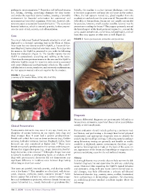
You are an Advanced Tactical Paramedic attached to small unit FIGURE 2 Severe pyotraumatic dermatitis and pyoderma.
deployed to a forward operating base in the Horn of Africa.
Your team has one Operational K9 (OpK9), a 3-year-old Ger-
man Shepherd, but no attached veterinary assets. Four days into
the mission, the OpK9 is presented to you with the following
lesion for evaluation (Figure 1). The handler reports that the
OpK9 is compulsively scratching and rubbing at the lesion.
There is no known previous trauma to the area and the OpK9 is
otherwise healthy except for recurrent otitis externa associated
with yeast (Malassezia pachydermatis) infections. The superfi-
cial skin lesion is moist, exudative, and extremely erythematous;
the hair appears matted and stuck together by the exudate.
FIGURE 1 Dog with lesion
(courtesy of Dr Amelia White, DVM, MS, DACVD).
Diagnosis
Primary differential diagnoses are pyotraumatic folliculitis or
furunculosis; dermatitis, superficial (bacterial or yeast [Malas-
Clinical Presentation sezia]); or dermatophytosis.
Pyotraumatic dermatitis may occur in any age, breed, sex, or Patient evaluation should include gathering a pertinent med-
discipline of canine; however, in one report, male dogs and ical history and performing a thorough head-to-tail physical
5
dogs younger than 4 years had a greater predisposition. examination and appropriate diagnostic tests to identify the
Breeds with a thick hair coat or dense undercoat (e.g., Golden underlying dermatologic etiology. Collecting samples and pur-
retriever, Labrador retriever, German Shepherd, Collie, Saint suing laboratory testing are generally not feasible or readily
1,5
Bernard) are also considered at higher risk. These same available in deployed, austere environment; therefore, a pre-
breeds are typically predisposed to the various underlying con- sumptive field diagnosis is made on the basis of the acute on-
ditions that triggers hot spots (e.g., atopic dermatitis, food al- set, appearance of the lesion and knowledge or suspicion of a
lergy dermatitis, otitis externa). Although a longer hair coat primary underlying cause for the self-inflicted trauma.
1,5
is typically touted as a predisposing factor, an analysis of 40
dogs with pyotraumatic dermatitis did not demonstrate any History
significant difference in incidence rates between dogs having A thorough history may provide clues to help narrow the dif-
either long or short hair coats. 6 ferential diagnosis list and determine the primary underlying
cause. Historical data regarding the OpK9’s typical behavior
Hot spots are painful, intensely pruritic, and develop rapidly and demeanor, along with any recent environmental and so-
1,2
over a few hours. They manifest as a localized, well-demar- cial changes, may help differentiate a primary self-directed
cated, alopecic, erythemic, moist, exudative wound. Acute behavioral disorder (e.g., anxiety, stress, conflict, frustration)
2,5
lesions appear flat and tend to have a central surface coagu- from a physiologic or dermatologic condition. For discerning
lum and erythematous perimeter, whereas chronic lesions may an underlying primary medical condition inquire about the
appear lichenified (i.e., raised, thickened) or granuloma-like. following:
1
106 | JSOM Volume 18, Edition 2/Summer 2018