
Page 97 - JSOM Winter 2017
P. 97
This can be known as extension lag and one finger may appear
not to fully extend. To assess this tendon, the affected finger
should be extended (Figure 3). If no lag is noted, then the re-
sistance should be applied to the finger to determine strength
against resistance and assess if pain occurs.
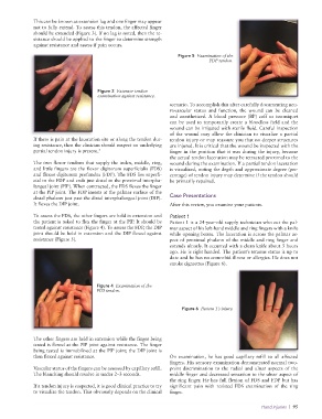
Figure 5 Examination of the
FDP tendon.
Figure 3 Extensor tendon
examination against resistance.
scenario. To accomplish this after carefully documenting neu-
rovascular status and function, the wound can be cleaned
and anesthetized. A blood pressure (BP) cuff or tourniquet
can be used to temporarily create a bloodless field and the
wound can be irrigated with sterile fluid. Careful inspection
of the wound may allow the clinician to visualize a partial
If there is pain at the laceration site or along the tendon dur- tendon injury or may reassure you that no deeper structures
ing resistance, then the clinician should suspect an underlying are injured. It is critical that the wound be inspected with the
partial tendon injury is present. 2 finger in the position that it was during the injury, because
the actual tendon laceration may be retracted proximal to the
The two flexor tendons that supply the index, middle, ring, wound during the examination. If a partial tendon laceration
and little fingers are the flexor digitorum superficialis (FDS) is visualized, noting the depth and approximate degree (per-
and flexor digitorum profundus (FDP). The FDS lies superfi- centage) of tendon injury may determine if the tendon should
cial to the FDP and ends just distal to the proximal interpha- be primarily repaired.
langeal joint (PIP). When contracted, the FDS flexes the finger
at the PIP joint. The FDP inserts at the palmar surface of the
distal phalanx just past the distal interphalangeal joint (DIP). Case Presentations
It flexes the DIP joint. After this review, you examine your patients.
To assess the FDS, the other fingers are held in extension and Patient 1
the patient is asked to flex the finger at the PIP. It should be Patient 1 is a 24-year-old supply technician who cut the pal-
tested against resistance (Figure 4). To assess the FDP, the DIP mar aspect of his left-hand middle and ring fingers with a knife
joint should be held in extension and the DIP flexed against while opening boxes. The laceration is across the palmar as-
resistance (Figure 5). pect of proximal phalanx of the middle and ring finger and
extends ulnarly. It occurred with a clean knife about 3 hours
ago. He is right handed. The patient’s tetanus status is up to
date and he has no comorbid illness or allergies. He does not
smoke cigarettes (Figure 6).
Figure 4 Examination of the
FDS tendon.
Figure 6 Patient 1’s injury.
The other fingers are held in extension while the finger being
tested is flexed at the PIP joint against resistance. The finger
being tested is immobilized at the PIP joint; the DIP joint is
then flexed against resistance. On examination, he has good capillary refill to all affected
fingers. His sensory examination demonstrated normal two-
Vascular status of the fingers can be assessed by capillary refill. point discrimination to the radial and ulnar aspects of the
The blanching should resolve in under 2–3 seconds. middle finger and decreased sensation to the ulnar aspect of
the ring finger. He has full flexion of FDS and FDP but has
If a tendon injury is suspected, it is good clinical practice to try significant pain with resisted FDS examination of the ring
to visualize the tendon. This obviously depends on the clinical finger.
Hand Injuries | 95