
Page 96 - JSOM Winter 2017
P. 96
Tetanus and Immune System Status Table 1 Neurological Examinations of the Fingers
Part of good wound management is understanding the pa- Nerve Sensory Examination Motor Examination
tient’s tetanus status and risk, and their immune system status. Radial Dorsal thumb–index Thumb extension
Certain medical conditions could affect the patient’s ability to finger web space
fight off infection. Patients with diabetes and tobacco use are Ulnar Palmar tip of the little Resisted abduction of the
particularly at risk for delayed healing and infection. 1 finger fingers
Median Palmar surface of index Resisted thumb palmar
finger abduction
Examination
Examination of the hand should be conducted systematically
after gaining a clear understanding of the mechanism of injury.
It is important to have know the underlying anatomy of the
hand and wrist. A good hand examination includes assessment
of neurological sensation and function, tendon examination,
vascular assessment, range of motion, and palpation.
Figure 2 Two-point discrimination.
A full anatomical review is beyond the scope of this article,
but we will review a few key features, especially as applied to
the fingers.
Nomenclature
When describing injuries to the hand it is important to use
concise and reliable terms that your colleagues can under-
stand. This is critical for the tactical clinician because you Bony Structures and Soft Tissue
may have to describe the injury to a higher medical author-
ity without the benefit of time or good clear communications. Bones of the Hand
Generally, the back of the hand is the dorsum (or the dorsal Moving distally from the wrist, the carpometacarpal joints are
aspect) and the palm side is referred to as palmar (or volar). formed between the carpels and metacarpals. The metacarpals
The terms medial and lateral can be indistinct, so the terms then extend the length of the palm and meet the phalanges at
radial (thumb side) or ulnar (little-finger side) are used for lo- the metacarpophalangeal joints. Each individual digit is com-
calization. There are many naming systems for the fingers, but posed of three segments of phalange, with the thumb being the
it is most accurate and simple to name them as thumb, index, exception as it is composed of only two phalange segments.
middle, ring, and little finger. Intersections of the phalanges form the proximal interphalan-
geal and distal interphalangeal joints, respectively. When per-
Neurological forming an assessment, taking note of the general appearance
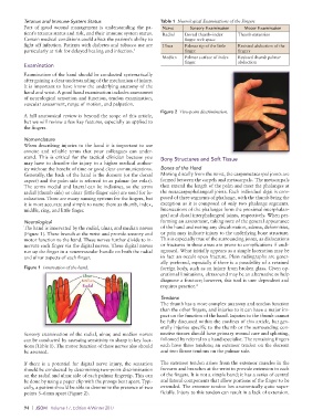
The hand is innervated by the radial, ulnar, and median nerves of the hand and noting any discoloration, edema, deformities,
(Figure 1). These branch at the wrist and provide sensory and or pain may indicate injury to the underlying bone structure.
motor function to the hand. These nerves further divide to in- This is especially true of the surrounding joints, as dislocations
nervate each finger via the digital nerves. These digital nerves or fractures in these areas are prone to complications if undi-
run up the finger in a neurovascular bundle on both the radial agnosed. What initially appears as a simple laceration may be
and ulnar aspects of each finger. in fact an occult open fracture. Plain radiographs are gener-
ally preferred, especially if there is a possibility of a retained
Figure 1 Innervation of the hand. foreign body, such as an injury from broken glass. Given op-
erational limitations, ultrasound may be an alternative to help
diagnose a fracture; however, this tool is user dependent and
requires practice. 2
Tendons
The thumb has a more complex anatomy and tendon function
than the other fingers, and injuries to it can have a major im-
pact on the function of the hand. Injuries to the thumb cannot
be fully discussed within the confines of this article, but gen-
erally injuries specific to the thumb or the surrounding con-
Sensory examination of the radial, ulnar, and median nerves nective tissues should have primary wound care and splinting,
can be conducted by assessing sensitivity to sharp in key loca- followed by referral to a hand specialist. The remaining fingers
tions (Table 1). The motor function of these nerves also should each have three tendons; an extensor tendon on the dorsum
be assessed. and two flexor tendons on the palmar side.
If there is a potential for digital nerve injury, the sensation The extensor tendon arises from the extensor muscles in the
should be conducted by determining two-point discrimination forearm and branches at the wrist to provide extension to each
on the radial and ulnar side of each palmar fingertip. This can of the fingers. It is not a simple band; it has a series of central
be done by using a paper clip with the prongs bent apart. Typi- and lateral components that allow portions of the finger to be
cally, a patient should be able to determine the presence of two extended. The extensor tendon lies anatomically quite super-
points 5–6mm apart (Figure 2). ficially. Injury to this tendon can result in a lack of extension.
94 | JSOM Volume 17, Edition 4/Winter 2017