
Page 68 - Journal of Special Operations Medicine - Fall 2017
P. 68
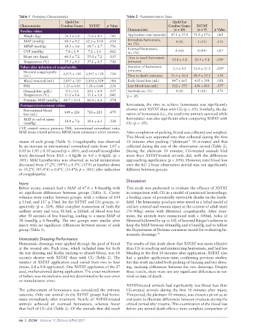
Table 1  Preinjury Characteristics                 Table 2  Postintervention Data
                                QuikClot                                           QuikClot
           Characteristic     Combat Gauze  XSTAT   p Value                      Combat Gauze   XSTAT
           Baseline values                                    Characteristic       (n = 10)    (n = 9)  p Value
             Weight (kg)        76.3 ± 5.0  75.6 ± 4.1  .745  Application time (seconds)  87.1 ± 17.4  71.5 ± 17.6  .069
             MAP (mmHg)         60.4 ± 9.2  62.5 ± 10.4  .654  Immediate hemostasis,    0 (0)  1 (11)   .474
                                                              no. (%)
             MPAP (mmHg)        18.3 ± 1.6  18.7 ± 2.7  .756  Eventual hemostasis,
             CVP (mmHg)         7.4 ± 1.9  7.2 ± 1.6  .862    no. (%)               4 (40)     8 (89)   .057
             Heart rate (bpm)   60.3 ± 7.2  55.0 ± 7.2  .135  Time to reach hemostasis                     a
             Temperature (°C)   37.3 ± 0.3  37.1 ± 0.7  .710  (minutes)            33.8 ± 4.8  20.3 ± 9.8  .028
           Values after induction of coagulopathy             Duration of hemostasis   5.4 ± 9.5  25.6 ± 31.3  .029 a
              Hextend coagulopathy   2,975 ± 192  2,947 ± 159  .728  (minutes)
            (mL)                                              Time to death (minutes)  35.4 ± 16.0  48.9 ± 29.1  .438
             Blood removed (mL)  2,817 ± 187  2,852 ± 209  .704  Early blood loss (mL)   847 ± 665  435 ± 398  .058
             INR               1.25 ± 0.05  1.28 ± 0.04  .228  Late blood loss (mL)  312 ± 373  434 ± 435  .377
              Hemoglobin (g/dL)   9.9 ± 0.6  10.1 ± 0.9  .557  Survival, no. (%)     0 (0)     1 (11)   .474
            Temperature (°C)    35.2 ± 0.6  35.1 ± 1.0  .841  a p < .05.
              Preinjury MAP (mmHg)  68.7 ± 10.1  66.4 ± 6.4  .574
           Postinjury/pretreatment values                    h emostasis, the time to achieve hemostasis was significantly
              Pretreatment blood    849 ± 228  720 ± 225  .479  shorter with XSTAT than with CG (p < .05). Similarly, the du-
            loss (mL)                                        ration of hemostasis (i.e., the total time animals survived while
              MAP at end of injury                           hemostatic) was also significant when comparing XSTAT with
            (mmHg)              38.9 ± 7.6  38.1 ± 6.4  .538  CG (p < .05).
          CVP, central  venous pressure;  INR, international normalized  ratio;
          MAP, mean arterial pressure; MPAP, mean pulmonary artery pressure.  After completion of packing, blood was collected and weighed.
                                                             This blood was separated into that collected during the first
          means of each group (Table 1). Coagulopathy was observed   10 minutes after packing (“platinum” 10 minutes) and that
          by an increase in international normalized ratio from 1.07 ±   collected during the rest of the observation period (Table 2).
          0.05 to 1.45 ± 0.10 second (p < .001), and overall hemoglobin   During the platinum 10 minutes, CG-treated animals bled
          levels decreased from 10.0 ± 0.8g/dL to 4.0 ± 0.4g/dL (p <   more than XSTAT-treated animals did, with the differences
          .001). Mild hypothermia was observed as rectal temperature   approaching significance (p = .058). However, total blood loss
          decreased from 37.2°C (99°F) ± 0.5°C (33°F) at baseline down   over the full 2-hour observation period was not significantly
          to 35.2°C (95.4°F) ± 0.8°C (33.4°F; p < .001) after induction   different between groups.
          of coagulopathy.
                                                             Discussion
          Injury
          Before injury, animals had a MAP of 67.6 ± 8.4mmHg with   This study was performed to evaluate the efficacy of XSTAT
          no  significant differences  between  groups  (Table  1).  Cavity   in comparison with CG in a model of junctional hemorrhage,
          volumes were similar between groups, with a volume of 104   a leading cause of potentially survivable deaths on the battle-
          ± 15mL and 117 ± 33mL for the XSTAT and CG groups, re-  field. The hemostatic products were tested in a lethal model of
          spectively (p = .324). After complete transection of both the   axillary arterial and venous injury in the context of adult-sized
          axillary artery and vein, 813mL ± 220mL of blood was lost   (70–90kg) swine with dilutional coagulopathy. After treat-
          after 30 seconds of free bleeding, leading to a mean MAP of   ment, the animals were resuscitated with a 500mL bolus of
          38.5mmHg ± 6.9mmHg. The two groups were similar after   Hextend followed by up to 10L of lactated Ringer’s solution to
          injury with no significant differences between means of each   keep the MAP between 60mmHg and 65mmHg and to follow
          group (Table 1).                                   the Department of Defense consensus model for evaluating he-
                                                             mostatic dressings. 18
          Hemostatic Dressing Performance
          Hemostatic dressings were applied through the pool of blood   The results of this study show that XSTAT was more effective
          at the wound site. Pack time, which included time for both   than CG in reaching and maintaining hemostasis, and had less
          the test dressing and Kerlix backing to absorb blood, was 16   bleeding in the first 10 minutes after application. XSTAT also
          seconds  shorter  with  XSTAT  than  with  CG (Table  2). The   had a quicker application time, confirming previous studies,
          number of XSTAT applicators used varied from two to four   but this study included both packing of backing and test dress-
          (mean, 2.8 ± 0.8 applicator). One XSTAT applicator, of the 27   ing, masking differences between the two dressings. Despite
          used, malfunctioned during application. The exact mechanism   these results, there were not any significant differences in sur-
          of failure was inconclusive and not determined to be user error   vival or time of death.
          or manufacturer error.
                                                             XSTAT-treated animals had significantly less blood loss than
          The achievement of hemostasis was considered the primary   CG-treated animals during the first 10 minutes after injury.
          outcome. Only one animal (in the XSTAT group) had hemo-  This period, the platinum 10 minutes, was chosen a priori as an
          stasis immediately after treatment. Nearly all XSTAT-treated   end point to illustrate differences between products during the
          animals  achieved  an  eventual  hemostasis,  whereas  fewer   critical period after trauma. This examination of the blood loss
          than half of CG did (Table 2). Of the animals that did reach   before any animal death offers a more complete comparison of

          66  |  JSOM   Volume 17, Edition 3/Fall 2017