
Page 67 - Journal of Special Operations Medicine - Fall 2017
P. 67
before treatment or a significant deviation from protocol oc- The time it took to pack both the test product and Kerlix
curred. An overview of the experimental protocol is shown in backing was defined as pack time. Hemostasis was defined as
Figure 1. no blood leaving the wound cavity, and immediate hemostasis
was defined as hemostasis occurring at the end of wound pack-
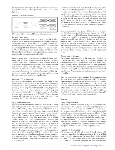
Figure 1 Experimental schematic. ing. Duration of hemostasis is the time during the experiment
when hemostasis was occurring while the animal was alive.
Blood flowing from the wound was collected by suction after
treatment in two phases: the initial 10 minutes (early blood
loss) and the remainder of the 2-hour observation period (late
blood loss).
After injury, animals were given a 500mL bolus of Hextend
at 100mL/min through the left external jugular vein. Follow-
EBV, estimated blood volume; LRS, lactated Ringer’s solution.
ing this bolus, up to 10L of lactated Ringer’s solution was ad-
Surgical Preparation ministered at 100mL/min to maintain a mean arterial pressure
Animals were fasted overnight before surgery but allowed free between 60mmHg and 65mmHg, in keeping with previous
access to water. They were sedated with 4.4mg/kg tiletamine– similar studies. Death was defined as a mean arterial pres-
18
zolazepam intramuscularly (IM) and 2.2mg/kg ketamine IM. sure (MAP) less than 20mmHg and end-tidal carbon dioxide
Buprenorphine was then given for alleviation of pain at a dose (Etco ) less than 15mmHg maintained for 2 minutes. Animals
2
of 0.01mg/kg IM. Anesthesia was induced via mask with 2% were killed by an overdose of pentobarbital once death crite-
to 4% isoflurane in an air/oxygen mixture of 40% to 60%. ria were reached or when 2 hours had passed from the initial
After intubation, isoflurane was adjusted to maintain a mini- injury.
mum alveolar concentration of at least 1.2.
Outcomes and Analysis
Vascular access was obtained using a modified Seldinger tech- The primary outcomes used in this study were survival, he-
nique. The left external jugular vein was accessed for resus- mostasis, and blood loss. Secondary outcomes included the
citation fluids, and a pulmonary artery catheter (Edwards following hemodynamic parameters: heart rate, MAP, Etco ,
2
Lifesciences, http://www.edwards.com) was inserted via the cardiac output, central venous pressure, and mean pulmonary
right external jugular vein. The right carotid artery was ac- artery pressure. Metabolic factors analyzed included lactate
cessed to monitor blood pressure and to allow for blood level, base excess, pH, and, the volume of resuscitation fluids
sampling. Splenectomy was performed through a midline lap- (i.e., lactated Ringer’s solution) used to maintain MAP above
arotomy to prevent splenic autoperfusion during hemorrhage, 60mmHg.
followed by a cystostomy for urine collection.
Data are presented as mean ± standard deviation unless other-
Induction of Coagulopathy wise noted. One-way analysis of variance (ANOVA) was used
Induction of coagulopathy was performed according to pre- for most analyses. However, Kruskal-Wallis ANOVA on ranks
vious studies. 5,17 Briefly, 60% of the estimated blood volume was used when the normality test failed (i.e., if p < .05). Sur-
was removed via the right femoral artery at 50mL/min. Simul- vival and hemostasis were analyzed using the Fisher exact test.
taneously, room-temperature Hextend (BioTime, http://www Additionally, survival was analyzed by log-rank analysis. Sta-
.biotimeinc.com) was infused at the same rate through the tistical analysis and data management were performed using
right-side external jugular vein. Hypothermia was allowed to Excel 2010 (Microsoft, www.microsoft.com) and Sigmaplot
progress until a temperature of 34.5°C (94.1°F) was reached; 12 (Systat Software, https://systatsoftware.com).
subsequently, warming blankets were used to keep tempera-
tures near 34.5°C (94.1°F) until the injury phase.
Results
Injury and Intervention Study Group Statistics
To gain access to the axillary artery and vein, a 4cm incision A total of 19 animals weighing 75.9kg ± 4.5kg were included
was made parallel to the sternum over the pectoralis major for analysis in this study and randomly assigned to groups as
muscle. The axillary artery, axillary vein, and brachial plexus follows: CG (n = 10 animals) and XSTAT (n = 9 animals). The
were then minimally dissected away (approximately 2cm) two groups were similar at baseline with no statistically signifi-
from the surrounding tissue. Wound cavity volume was de- cant differences between means of each group (Table 1). Four
termined by measuring the amount of warmed saline neces- animals had to be excluded from the analysis: one animal died
sary to fill the wound cavity. The vessels were then bathed in during coagulopathy and was not included in randomization,
2% lidocaine for 10 minutes to induce dilation. After suction one animal from the CG group and one from the XSTAT group
removal of lidocaine, a necropsy blade was used to transect were excluded because of a protocol deviation of 45% oxygen
both the axillary artery and vein to initiate injury (time = 0 during injury, and one animal randomly assigned to the XSTAT
minutes). Hemorrhage was allowed to proceed for 30 seconds group was excluded because of infusion pump failure.
while blood was collected by suction and weighed. The test he-
mostatic dressing was then applied to the wound using either a Induction of Coagulopathy
single roll of CG or up to four XSTAT applicators. Kerlix was The replacement of 60% of estimated blood volume with
packed into the wound as backing, but no manual compres- Hextend resulted in the administration of 2,962 ± 172mL
sion was applied in either group. This deviation from both over 59.2 ± 3.5 minutes and the removal of 2,794 ± 162mL
products’ instructions for use was done to test them as equivo- of blood. The two groups were similar after coagulopathy
cally as possible. induction and there were no significant differences between
Junctional Hemorrhage Model Testing XSTAT | 65
®