
Page 114 - Journal of Special Operations Medicine - Spring 2017
P. 114
An Ongoing Series
Meningococcal Disease
Mark W. Burnett, MD
Introduction to evaluate in young children and infants. Meningo-
coccemia, or bloodstream infections, may occur with
It’s a not uncommon story in the newspaper—college or without meningitis and have features of limb pain,
student goes to bed with “flu-like symptoms” only to purpura, shock, adrenal hemorrhage, and multisystem
be found dead the next day after not reporting for class. organ failure. In both meningococcal meningitis and
Meningococcal disease, caused by the bacterium Neis- sepsis, initial symptoms may be “flu-like,” which may
seria meningitidis, is one of the most common causes of lead to a delay in the patient seeking care. Less common
sepsis and meningitis in the United States and a feared presentations of this disease include pneumonia (up to
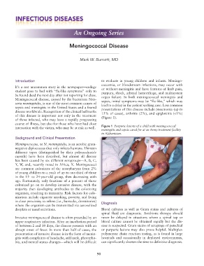
disease worldwide. Recognition of the clinical hallmarks 15% of cases), arthritis (2%), and epiglottitis (<1%)
of this disease is important not only in the treatment (Figure 1).
of those infected, who may have a rapidly progressing
course of illness, but also for those who have had close
interaction with the victim, who may be at risk as well. Figure 1 Purpuric lesions of a child with meningococcal
meningitis and sepsis cared for at an Army treatment facility
in Afghanistan.
Background and Clinical Presentation
Meningococcus, or N. meningitidis, is an aerobic gram-
negative diplococcus that only infects humans. Thirteen
different types (distinguished by their polysaccharide
capsule) have been described, but almost all disease
has been caused by six different serogroups—A, B, C,
Y, W, and, recently noted in Africa, X. Meningococci
are common colonizers of the nasopharynx from 2%
of young children to a peak of up to one-third of those
in the 15- to 24-year-old group, then decreasing with
age. Fortunately, only fractions of a percent of those
colonized go on to develop invasive disease, with the
majority then developing antibodies to the colonizing
organism, resulting in immunity. Risk factors for colo-
nization include cigarette smoking, poverty, and living
in close proximity to others (i.e., barracks, dormitories) Diagnosis
where the organism can be transmitted via aerosolized
droplets or nasal secretions. Blood cultures as well as Gram stains and cultures of
spinal fluid are diagnostic. Antibiotic therapy should
Invasive meningococcal disease is often preceded by an never be delayed in situations where a spinal tap or
upper respiratory infection. After an incubation period blood culture cannot be obtained rapidly but the dis-
of between 2 and 10 days, the disease presents with an ease is suspected. Gram stains of scrapings of petechial
abrupt onset of fever. In more than half of cases, the or purpuric lesions may also prove helpful. Multiplex
presentation of invasive disease is in the form of menin- polymerase chain reaction testing, as is found in large
gitis with complaints of headache, stiff neck, photopho- hospitals and occasionally in deployed environments,
bia, and mental status changes—which will be difficult can significantly shorten the time to definitive diagnosis.
90