
Page 23 - Journal of Special Operations Medicine - Spring 2016
P. 23
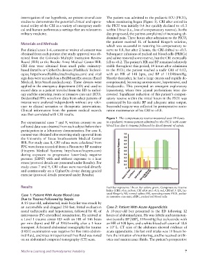
interrogation of our hypothesis, we present several case   The patient was admitted to the pediatric ICU (PICU),
              studies to demonstrate the potential clinical and opera-  where monitoring began (Figure 1). CRI after arrival in
              tional utility of the CRI in a variety of emergency medi-  the PICU was initially 0.4 but quickly declined to <0.3
              cal and human performance settings that are relevant to   within 1 hour (i.e., loss of compensatory reserve). As the
              military medicine.                                 day progressed, the patient complained of increasing ab-
                                                                 dominal pain. Three hours after admission to the PICU,
                                                                 the patient received 1L of lactated Ringer’s solution,
              Materials and Methods
                                                                 which was successful in restoring his compensatory re-
              For clinical cases 1–6, consent or waiver of consent was   serve to 0.8, but after 2 hours, the CRI drifted to <0.3.
              obtained from each patient after study approval was ob-  Subsequent infusions of packed red blood cells (PRBCs)
              tained from the Colorado Multiple Institution Review   and saline restored some reserve, but the CRI eventually
              Board (IRB) or the Brooke Army Medical Center IRB.   fell to <0.2. The patient’s HR and BP remained relatively
              CRI data were obtained from small pulse oximetry-  stable throughout this period; 14 hours after admission
              based DataOx or CipherOx devices (Flashback Technol-  to the PICU, the patient reached a nadir CRI of 0.12,
              ogies; http://www.flashbacktechnologies.com) and vital   with an HR of 148 bpm, and BP of 115/48mmHg.
              sign data were recorded on a BedMasterEx system (Excel   Shortly thereafter, he had a large emesis and rapidly de-
              Medical; http://excel-medical.com). These devices were   compensated, becoming unresponsive, hypotensive, and
              applied in the emergency department (ED) and used to   bradycardic. This prompted an emergent exploratory
              record data as a patient traveled from the ED to radiol-  laparotomy, where two jejunal perforations were dis-
              ogy and the operating room or intensive care unit (ICU).   covered. Significant reduction of this patient’s compen-
              Deidentified PPG waveform data from select patients of   satory reserve within the initial hours in the PICU was
              interest were analyzed independently without any refer-  contrasted by his stable BP and adequate urine output.
              ence  to clinical  scenarios or  therapeutic interventions.   Successful surgery was reflected by postoperative resto-
              Clinical  information  from the patient  medical records   ration maintenance of his CRI to >0.8.
              was then correlated with CRI results.
                                                                 Figure 1  The compensatory reserve measured over 29 hours
              For experimental cases 7 and 9, written consent to use   in a pediatric trauma patient admitted to the PICU with acute
              collected data was obtained from each subject before their   blood loss due to trauma followed by development of sepsis.
              participation in a laboratory demonstration. For case 8,
              consent was obtained after receiving study approval from
              the University of Texas Southwestern Medical Center
              IRB. For study case 8, CRI values were calculated from
              PPG waveforms recorded from a Finometer BP monitor
              (Finapres Medical Systems; http://www.finapres.com)
              during exposure to progressive lower-body negative
              pressure (LBNP) with and without exposure to a heat
              stress (protocol details are presented under Results). For
              study cases 7 and 9, CRI values were recorded directly
              and continuously on a CipherOx device during graded
              exercise (protocol details presented under Results).


              Results                                            Each bar represents 1 hour. Bar colors: green, Compensatory Reserve
                                                                 Index (CRI) >0.6; yellow, CRI ≤0.6 and >0.3; red, CRI ≤0.3. LR, lac-
              Case 1: Patient With Acute Blood Loss              tated Ringer’s; NS, normal saline; OR, operating room; PICU, pediat-
                                                                 ric intensive care unit; pRBC, packed red blood cells.
              Due to Trauma Followed by Sepsis
              A 15-year-old, unhelmeted, male bicyclist was struck by
              an automobile and dragged 250 feet. Initial evaluation   Case 2: Patient With Acute Appendicitis
              noted tachycardia and hypotension, followed by 4L of   A 10-year-old boy presented to the ED following 12
              intravenous (IV) crystalloid resuscitation. He arrived at   hours of abdominal pain. He was febrile and normoten-
              a Level I trauma center ED with an HR of 146 beats   sive (systolic BP [SBP], 118mmHg) but tachycardic with
              per min (bpm) and BP of 99/61mmHg after a 1-hour   an HR of 108 bpm, and a white blood cell count of 18.6
              transport. A focused abdominal sonography for trauma   × 10  L. CT scan of the abdomen showed evidence of
                                                                     9
              (FAST) examination was negative for free intra-abdom-  acute appendicitis. His last oral intake was 10 hours be-
              inal fluid, and trace intraperitoneal free fluid was noted   fore the initiation of monitoring, and he received antibi-
              on an abdominal computed tomography (CT) scan.     otics and maintenance fluids. The patient’s preoperative


              Machine Learning and Hemodynamic Instability                                                     7