
Page 41 - Journal of Special Operations Medicine - Winter 2014
P. 41
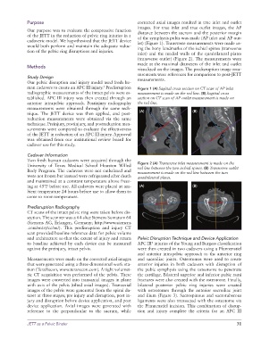
Purpose                                            corrected axial images resulted in true inlet and outlet
                                                                 images.  For true  inlet and true  outlet images,  the  AP
              Our purpose was to evaluate the compressive function   distance between the sacrum and the posterior margin
              of the JETT in the reduction of pelvic ring injuries in a   of the symphysis pubis was made (AP inlet and AP out-
              cadaveric model. We hypothesized that the JETT device   let) (Figure 1). Transverse measurements were made us-
              would both perform and maintain the adequate reduc-  ing the bony landmarks of the ischial spines (transverse
              tion of the pelvic ring disruptions and injuries.
                                                                 inlet) and the medial walls of the quadrilateral plates
                                                                 (transverse outlet) (Figure 2). The measurements were
                                                                 made at the maximal diameters of the inlet and outlet
              Methods
                                                                 visualized on the images. The predisruption image mea-
                                                                 surements were references for comparison to post-JETT
              Study Design                                       measurements.
              Our pelvic disruption and injury model used fresh hu-
              man cadavers to create an APC III injury.  Predisruption   Figure 1 (A) Sagittal cross section on CT scan of AP inlet
                                                 5
              radiographic measurements of the intact pelvis were es-  measurement is made on the red line. (B) Sagittal cross
              tablished. APC III injury was then created through an   section on CT scan of AP outlet measurement is made on
              anterior intrapelvic approach. Postinjury radiography   the red line.
              measurements were obtained through the same tech-
              nique. The JETT device was then applied, and post-  (A)                     (B)
              reduction measurements were obtained via the same
              technique. Preinjury, postinjury, and postreduction mea-
              surements were compared to evaluate the effectiveness
              of the JETT in reduction of an APC III injury. Approval
              was obtained from our institutional review board for
              cadaver use for this study.

              Cadaver Information
              Two fresh human cadavers were acquired through the
              University  of  Texas  Medical  School  Houston  Willed   Figure 2 (A) Transverse inlet measurement is made on the
                                                                 red line between the two ischial spines. (B) Transverse outlet
              Body Program. The cadavers were not embalmed and   measurement is made on the red line between the two
              were not frozen but instead were refrigerated after death   quadrilateral plates.
              and maintained at a constant temperature above freez-
              ing at 45ºF before use. All cadavers were placed at am-  (A)                (B)
              bient temperature 24 hours before use to allow them to
              come to room temperature.

              Predisruption Radiography
              CT scans of the intact pelvic ring were taken before dis-
              section. The scanner was a 64-slice Siemens Somatom 64
              (Siemens AG, Erlangen, Germany; http://www.siemens
              .com/entry/cc/en/). This predisruption and injury CT
              scan provided baseline reference data for pelvic volume
              and architecture so that the extent of injury and return   Pelvic Disruption Technique and Device Application
              to  baseline  achieved  by  each  device  can  be measured   APC III  injuries of the Young and Burgess classification
                                                                       5
              against the preinjury, intact pelvis.              were then created in two cadavers using a Pfannenstiel
                                                                 and anterior intrapelvic approach to the anterior ring
              Measurements were made on the corrected axial images   and sacroiliac joints.  Osteotomes were  used to create
              that were generated using a three-dimensional work sta-  anterior injuries in both cadavers with disruption of
              tion (TeraRecon, www.terarecon.com). A tight volumet-  the pubic symphysis using the osteotome to penetrate
              ric CT acquisition was performed of the pelvis. These   the cartilage. Bilateral superior and inferior pubic rami
              images were converted into transaxial images in plane   fractures were also created with the osteotome. Finally,
              with axis of the pelvis (tilted axial images). Transaxial   bilateral posterior pelvic ring injuries were created
              images of the pelvis were generated from the spiral da-  with osteotomes through the anterior sacroiliac joint
              taset at three stages; pre injury and disruption, post in-  and ilium (Figure 3). Sacrospinous and sacrotuberous
              jury and disruption before device application, and post   ligaments were also transected with the osteotome via
              device application. Axial images were generated with   the Pfannenstiel incision. This combination of disrup-
              reference  to the perpendicular to the sacrum, while   tion  and  injury  complete  the  criteria  for  an  APC  III


              JETT as a Pelvic Binder                                                                         31