
Page 56 - Journal of Special Operations Medicine - Spring 2014
P. 56
indentations on the sides for holding the device like a lateral to the blade tip are also designed to prevent ex-
pencil, with the second and third fingers on the indenta- cessive insertion.
tions. The clinician’s thumb is stabilized on a rounded
knob projecting up from the middle of the hook. The After the downward incision is made with the blade, the
hook is entirely made of brushed aluminum. thumb slides the hook down the handle into the hole
(by moving the knob and hook down the scalpel) (Fig-
The scalpel can be used in either a vertical or a horizon- ure 6). It should be advanced fully into hole. This is felt
tal orientation; in most emergent situations, it is advised mechanically as the leading edge of the hook passes over
to make a vertical skin incision, confirm the location of the scalpel and under the inferior edge of the thyroid
the cricothyroid membrane by palpation, and then ori- cartilage. The hook has a tip with a triangular shape;
ent the scalpel to a horizontal orientation for incising as it is advanced into the hole, there is a distinct click
the membrane. When the location of the cricothyroid once the hook passes under the thyroid cartilage. The
membrane is obvious, the blade can be inserted through hook slides over the scalpel and is kept flush along the
skin and membrane together (horizontally). Whether scalpel by the lateral projections of the hook, which
an initial vertical or horizontal incision is made on the move within a channel on the handle. Once the hook
skin, the cricothyroid membrane is always entered hori- has been advanced down the handle (so that it has ad-
zontally with a downward motion (perpendicular to the vanced fully into the hole made by the scalpel), the hook
membrane) aiming at the back wall of the cricoid car- can be lifted away from the scalpel and handle. The
tilage (Figure 5). It is important to note that the cricoid transition is quick and easy between the dominant hand
cartilage has much larger back wall than its relatively sliding the hook down (the right thumb in the photo-
small anterior ring. Unlike an incision at the level of the graphs) and the nondominant hand (the left hand in the
tracheal rings, where the back of the tracheal is soft, photographs) grabbing the hook once it has been fully
flat, and abuts the esophagus, the large back wall of the advanced. The hook has a large proximal finger hold
cricoid provides a firm protective stop. Also, because of that does not require fine motor control. The clinician
the tight fit of the cricoid cartilage within the overlying now has control of the trachea with the hook, and a
thyroid, there is also a cartilaginous stop at the lateral sufficiently large incision made by the scalpel, to allow
ends of the cricothyroid membrane. This prevents ex- insertion of the Cric-Key and overlying tube.
tension of an incision at the cricothyroid membrane too
far laterally, limiting the risk of injuring the carotid and The slide of the hook over the scalpel immediately into
jugular vessels. The Cric-Knife blade is short in length the hole reduces the risk of losing the hole; the tip of the
to prevent overinsertion (so as not to hit the posterior hook enters fully with a palpable click as it wedges un-
cricoid wall or the vocal cords), and plastic projections der the thyroid. The shape of the handle, the hook, and
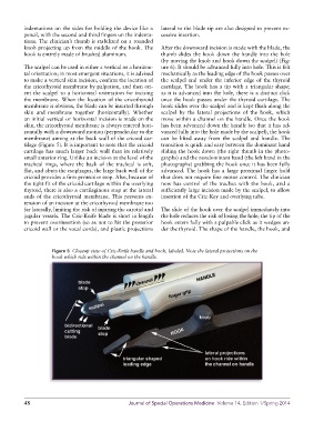
Figure 5 Closeup view of Cric-Knife handle and hook, labeled. Note the lateral projections on the
hook which ride within the channel on the handle.
48 Journal of Special Operations Medicine Volume 14, Edition 1/Spring 2014