
Page 116 - JSOM Fall 2025
P. 116
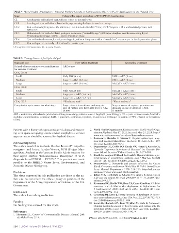
TABLE 2  Hydatid Cyst Symptomology by Anatomic Site  TABLE 3  Hydatid Cyst Differential Diagnosis by Anatomic Site
              Site                        Symptomology           Site                     Differential diagnoses
              Liver 2            Abdominal pain                  Liver 2            Benign tumor
                                 Nausea                                             Malignancy
                                 Vomiting                                           Amebic abscess
                                 Feeling of pressure                                Congenital cyst
                                 Jaundice 7                      Lungs              Tuberculosis
              Lungs 2            Chronic cough                                      Carcinoma
                                 Chest pain                                         Fungal infection
                                 Dyspnea                         Heart 5            Congenital cyst
              Heart 5            Precordial discomfort                              Aneurysm
                                 Palpitation                                        Fibroma
                                 Dyspnea                                            Myxoma
                                 Arrhythmia                      Bone 5             Tuberculous spondylitis
                                 Cardiac tamponade                                  Chronic osteomyelitis
                                 Acute pericarditis                                 Neurofibroma
                                 Anaphylaxis                                        Metastases
              Bone 5             Localized pain                                     Giant cell tumor
                                 Destruction of bone cortex                         Meningocele
              Orbit 5            Unilateral painless proptosis                      Bone cyst
                                 Orbital pain                                       Developmental cyst
                                 Chemosis                        Orbit 5            Abscess
                                 Orbital cellulitis                                 Intraorbital hematoma
                                 Loss of vision                                     Lacrimal tumor
              Mediastinum 5      Dyspnea                                            Mucocele
                                 Cough                                              Lymphangioma
                                 Retrosternal chest pain         Mediastinum 5      Meningocele
                                 Dysphagia                                          Mature cystic teratoma
                                 Dysphasia                                          Thymic lymphangioma
                                 Back pain                                          Thymoma
                                 Superior vena cava syndrome                        Mediastinal carcinoma
                                 Mediastinal widening                               Benign mass
              Kidney 5           Flank discomfort                Kidney 5           Cystic nephroma
                                 Lumbar mass                                        Simple renal cyst
                                 Abdominal mass                                     Necrotic renal cell carcinoma
                                 Subcostal pain                                     Renal abscess
                                 Vomiting                        Central nervous system 5  Brain abscess
                                 Fever                                              Intracranial arachnoid cyst
                                 Dysuria                                            Tumor
                                 Hematuria                       Spleen 5           Epidermoid cyst
              Central nervous system 5  Headache                                    Pseudocyst
                                 Vomiting                                           Solitary abscess
                                 Papilledema                                        Cystic tumor
                                 Peripheral neuropathy                              Hematoma
                                 Skull deformity                 Peritoneum         Perforated peptic ulcer
                                 Seizures                        (ruptured liver cyst)  Cholecystitis
              Spleen 5           Left hypochondrium mass                            Appendicitis
                                 Abdominal pain                                     Pancreatitis
                                 Fever                                              Ruptured sigmoid diverticulum
                                 Dyspnea                                            Ovarian torsion
                                 Dyspepsia                                          Volvulus
                                 Colon fistula                                      Ruptured abdominal aortic aneurysm
                                 Perforation of diaphragm
              Peritoneum         Anaphylaxis                     Dogs harboring E. granulosus tapeworms often appear to be
                      10
              (ruptured liver cyst)  Abdominal distension
                                 Diffuse tenderness              in good health, so the outward healthy appearance of a dog
                                 Diffuse dullness to percussion  should not be used as a gauge for the risk of hydatid disease
                                                                 transmission. While the servicemembers who contract cystic
                                                                 echinococcosis during a deployment will likely remain asymp-
              dog feces for gravid proglottids and/or eggs of E. granulosus.   tomatic during the deployment, it is probable they will suffer
              A vaccine (EG95) exists for prevention of the cystic stage in   deleterious effects from the steadily growing hydatid cyst in
              sheep, but it is only registered in China and Argentina. 2  years or decades. These effects include impingement of paren-
                                                                 chymal tissue with commensurate loss of function (specific to
                                                                 the cyst location) and/or anaphylactic death secondary to cyst
              Importance in a Deployed Setting
                                                                 rupture.  Within endemic regions, avoidance  of contact  with
              As discussed earlier, humans in physical contact with infected   dogs, fastidious handwashing, and thorough cleaning of food
              dogs put themselves in danger of contracting hydatid disease.   that may have come into contact with dog feces is paramount.

                                                                                                Hydatid Disease  |  113