
Page 61 - JSOM Summer 2025
P. 61
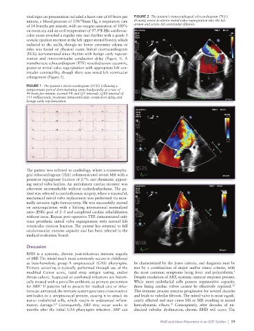
vital signs on presentation included a heart rate of 60 beats per   FIGURE 2  The patient’s transesophageal echocardiogram (TEE)
              minute, a blood pressure of 138/78mm Hg, a respiratory rate   showing severe posterior mitral valve regurgitation into the left
              of 14 breaths per minute, with an oxygen saturation of 100%   atrium and severe left ventricular dilation.
              on room air, and an oral temperature of 97.9°F. His cardiovas-
              cular exam revealed a regular rate and rhythm with a grade 3
              systolic ejection murmur at the left upper sternal border, which
              radiated to the axilla, though no lower extremity edema or
              rales was found on physical exam. Initial electrocardiogram
              (ECG) demonstrated sinus rhythm with benign early repolar-
              ization and intraventricular conduction delay (Figure 1).  A
              transthoracic echocardiogram (TTE) revealed severe eccentric,
              posterior mitral valve regurgitation with appropriate left ven-
              tricular contractility, though there was noted left ventricular
              enlargement (Figure 2).

              FIGURE 1  The patient’s electrocardiogram (ECG) following a
              symptomatic period demonstrating sinus bradycardia at a rate of
              44 beats per minute, normal PR and QT intervals, QRS interval of
              113 milliseconds, moderate intraventricular conduction delay, and
              benign early repolarization.


              The patient was referred to cardiology, where a transesopha-
              geal echocardiogram (TEE) redemonstrated severe MR with a
              posterior regurgitant fraction of 27% and rheumatic appear-
              ing mitral valve leaflets. An ambulatory cardiac monitor was
              otherwise unremarkable without tachydysrhythmia. The pa-
              tient was referred to cardiothoracic surgery, where a successful,
              mechanical mitral valve replacement was performed via mini-
              mally invasive right thoracotomy. He was successfully started
              on anticoagulation with a lifelong international normalized
              ratio (INR) goal of 2–3 and completed cardiac rehabilitation
              without issue. Repeat post-operative TTE demonstrated only
              trace prosthetic mitral valve regurgitation with normal left
              ventricular ejection fraction. The patient has returned to full
              cardiovascular exercise capacity and has been referred to the
              medical evaluation board.

              Discussion
              RHD is a systemic, chronic post-infectious immune sequela
              of ARF. The initial insult most commonly occurs in childhood
              as beta-hemolytic group A streptococcal (GAS) pharyngitis.   be characterized by the Jones criteria, and diagnosis may be
              Primary screening is typically performed through use of the   met by a combination of major and/or minor criteria, with
                                                                                                                5
              modified Centor score, rapid strep antigen testing, and/or   the most common symptoms being fever and polyarthritis.
              throat culture. Suspected or confirmed infections are histori-  Despite resolution of ARF, systemic immune response persists.
              cally treated with a penicillin antibiotic as primary prevention   While most endothelial cells possess regenerative  capacity,
                                                                                                               5,9
              for ARF.  If patients fail to present for medical care or infec-  those  lining  cardiac  valves  cannot  be  effectively  repaired.
                    7
              tions go untreated, the immune system generates cross-reactive   This immune process remains progressive for several decades
              antibodies to a streptococcal protein, causing it to attack its   and leads to valvular fibrosis. The mitral valve is most signifi-
              native endothelial cells, which results in widespread inflam-  cantly affected and may cause MS or MR resulting in mixed
                                                                                  10
              matory damage.  Consequently,  ARF may occur weeks to   hemodynamic  effects.  Consequently, after  decades of un-
                           8,9
              months after the initial GAS pharyngitis infection. ARF can   checked valvular dysfunction, chronic RHD will occur. The
                                                                             RHD and Valve Placement in an SOF Soldier  |  59