
Page 88 - JSOM Spring 2025
P. 88
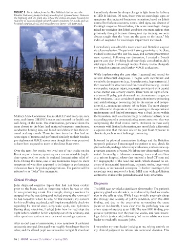
FIGURE 2  Photo taken from the Mar Mikhael district near the   immediately due to the abrupt change in light from the hallway
          Charles Helou highway, looking into the port (ground zero). Between   to LED-lit kitchen. Of note, there were no neurologic signs or
          the highway and the grain silo, where the cranes are, were located the   symptoms that indicated brainstem herniation, based on John’s
          majority of various supply wharehouses/containers for goods such as
          hospital supplies, food, and miscellaneous items—all destroyed.   normal level of consciousness, normal vital signs, and absence of
                                                             Cushing’s response. Nevertheless, this acute neurologic change
                                                             raised my suspicion that John’s condition was more serious than
                                                             previously thought because throughout my training, we were
                                                             always taught that the “eyes are the gates to the brain.” My
                                                             index of suspicion for neurologic impairment increased.

                                                             I immediately consulted the team leader and Battalion surgeon
                                                             via teleconsultation. The patient’s history, proximity to the blast,
                                                             medical events over the last few days, and current presentation
                                                             were reported. Following our discussion, I created a 2-week
                                                             patient care plan involving local neurologic consultation, daily
                                                             vital signs checks, a thorough medical history review alongside
                                                             my Battalion surgeon, and further MACE evaluations.

                                                             While implementing the care plan, I assessed and tested for
                                                             several differential diagnoses. I began with nutritional and
                                                             metabolic derangements (e.g., hypoglycemia, hyponatremia). I
                                                             then assessed for structural and functional lesions (e.g., cranial
                                                             nerve palsy, vascular injury, traumatic eye injury) with cranial
                                                             nerve, motor, and sensory exams. There were no signs of cra-
                                                             nial nerve III palsy, gait abnormalities, dermatome changes, or
             Photo by ZJL.                                   focal weakness. I also considered sympathomimetic poisoning
                                                             and anticholinergic poisoning due to the nature and compo-
                                                             nents (i.e., ammonium nitrate) of the blast. The most danger-
                                                             ous differential diagnosis at the time, owing to the operational
                                                             environment and limited resources, was a focal lesion within
                                          5
          Military Acute Concussion Exam (MACE)  and head, eye, ears,   the brainstem, such as a hemorrhage or ischemic infarct, or an
          nose, and throat (HEENT) exams and ensured the health and   expanding posterior communicating artery aneurysm that was
          well-being of the team. On examination, personnel from the   compressing the third cranial nerve. However, in those early
          team closest to the blast had ruptured tympanic membranes,   moments with limited information, my most likely differential
          conductive hearing loss, and blood and debris within their ex-  diagnosis was that this was related to post-blast exposure to
          ternal auditory canals. Those furthest from the blast had no   chemicals, such as anticholinergic poisoning.
          acute signs of trauma and performed similarly to their baseline
          pre-deployment MACE scores even though they were perceived   Informed by physical examination findings and the Battalion
          to have been exposed to more of the direct blast wave.  surgeon’s guidance, I encouraged the patient to rest, check his
                                                             glucose levels, undergo laboratory evaluation, and consume ap-
          Over the next few weeks, we lived out of our trucks on the   propriate amounts of water. No laboratory abnormalities were
          Beirut airport’s tarmac, operating on a reverse schedule (night-  noted. Eventually, a Lebanese neurology team evaluated him
          time operations) to assist in regional humanitarian relief ef-  at a private hospital, where they ordered a head CT scan and
          forts. During this time, one of my teammates began to show   CT angiography of the head and neck, which showed no evi-
          symptoms of what first appeared to be fatigue or performance   dence of intracranial hemorrhage, ischemic stroke, large vessel
          exhaustion from the prolonged operations. The patient will be   occlusion, or intracranial aneurysm. To further investigate, the
          referred to as “John” for anonymity.               neurology team requested a brain MRI scan with gadolinium
                                                             contrast to evaluate the parenchyma and bony structures.
          Clinical Findings
                                                             Diagnosis
          John displayed cognitive lapses that had not been evident
          prior to the blast, such as forgetting where he was or why   The MRI scan revealed a significant abnormality. The patient’s
          he was performing a task. For example, during a workout he   pituitary gland was shrunken, as evidenced by fluid accumula-
          stopped what he was doing and looked around the room as if   tion in the sella turcica. While I was initially uncertain about
          he had forgotten where he was. At that moment, my concern   the etiology and severity of John’s condition, after this MRI
          for his well-being escalated, and I implemented daily check-ins   finding, and due to the uncertainty surrounding the cause
          regarding his mental state, sleep-wake cycle, and hydration.   (acute or incidental), it was clear that his pathology was out
          During these check-ins, I asked John how he had slept the   of my scope of practice as an 18D. The new MRI finding, pro-
          night before, whether he felt anything out of the ordinary, and   gressive symptoms over the past few weeks, and focal neuro-
          other questions pertinent to a review of neurologic systems.  logic deficit (anisocoria) ultimately led me to advise our team
                                                             leader to medically evacuate John.
          After several days of assessments, a new symptom of unilateral
          anisocoria emerged. One pupil was roughly 4mm larger than the   I remember my team leader looking at me, relying entirely on
          other, and the dilated pupil was unreactive to light. It stood out   my clinical judgment to inform his command decision. This

          86  |  JSOM   Volume 25, Edition 1 / Spring 2025