
Page 72 - JSOM Fall 2023
P. 72
Pain Control and Point-of-Care Ultrasound
An Approach to Rib Fractures for the Austere Provider
1
Reece Snyder, PA-C *; Dan Brillhart, MD 2
ABSTRACT
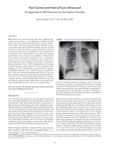
Rib fractures are common injuries that cause significant dis- FIGURE 1 Chest radiograph demonstrating initially read as normal.
comfort and can lead to severe pulmonary complications. Rib
injury most often results from high-velocity traumatic mecha-
nisms, while rarely representing underlying metastatic disease
or secondary injury due to pulmonary illness. Because most rib
fractures are caused by obvious trauma, algorithms are focused
on treatment rather than investigating the exact mechanism of
rib fractures. Chest radiographs are often the initial imaging
performed but have proven to be unreliable in identification
of rib fracture. Computed tomography (CT) is a diagnostic
option as it is more sensitive and specific than simple radio-
graphs. However, both modalities are generally unavailable to
Special Operations Forces (SOF) medical personnel working
in austere locations. These medical providers could potentially
diagnose and treat rib fractures in any environment using a
standardized approach that includes clarity of mechanism,
pain relief, and point-of-care ultrasound (POCUS). This case
demonstrates an approach to the diagnosis and treatment of a
rib fracture in a 47-year-old male who presented to a military
treatment facility with unlocalized flank and back pain, but
the methods employed have applicability to the austere pro-
vider working far from the resources of a medical center. assess for potential infectious, oncologic, renal, or bony ab-
normalities. Creatinine was modestly elevated but was similar
Keywords: pocus; rib fracture; perineural block; prolother- to bloodwork the patient had completed 2 months prior. After
apy; musculoskeletal ultrasound further chart evaluation, the patient had had an outpatient re-
nal ultrasound (US) the day prior for routine evaluation of
elevated creatinine. This ultrasound showed no abnormalities.
The chest radiograph was normal.
Background
A 47-year-old male presented to the Emergency Department Based on the above presentation, the patient was suspected to
(ED) of an Army Medical Center with the initial complaint of have an acute muscle strain secondary to his workout. Peri-
back pain for 4 days. The patient described a 4-day history of neural injections were performed by first identifying relevant
right posterior lateral thoracic pain after completing a rigor- landmarks including T11, the 11th rib, and the point of max-
ous leg workout that included the use of a belt squat machine. imal tenderness. Injections of 0.5mL of 1% lidocaine were
This was the first time the patient had used this machine, and then injected subcutaneously along the approximate courses
the patient stated he had recently increased the overall inten- of the cutaneous branch of the dorsal rami of T11 and the
sity of his lower body workouts. He decided to come to the lateral cutaneous branch of the 11th intercostal nerve. These
ED because the previous night he had sneezed and felt a “pop” injections were spaced 3-cm apart except around the point of
with increased pain. Physical exam showed full range of mo- maximal tenderness where the injections were spaced 1.5-cm
tion of the lumbar, thoracic, and cervical spine but moderate apart. Once analgesia was obtained, the area of maximal pain
tenderness along the paraspinous muscles of the right side be- (around T11) was palpated eliciting crepitus that was not pres-
tween T8 and T11 that radiated to the midaxillary line. ent on initial examination. POCUS was performed at bedside
(Figure 2) showing a mildly displaced fracture of the 11th rib.
Initial evaluation included a complete blood count and com- A CT scan with contrast was performed (Figure 3) in order
plete metabolic count with chest radiography (Figure 1) to to investigate potential solid organ injury. That CT confirmed
*Correspondence to reece.c.snyder@gmail.com
1 Reece Snyder is a recent graduate of the Interservice Physician Assistant Program, where he also completed the Emergency Ultrasound Intern-
ship. Dr Dan Brillhart is a physician and the fellowship director of the Emergency Ultrasound Fellowship, Carl R. Darnhall Army Medical
2
Center, Fort Cavazos, TX.
70