
Page 116 - JSOM Summer 2022
P. 116
the enzymatic formation of fibrin from fibrinogen. The loss FIGURE 4 Fibrinolysis.
of calcium from hemorrhage or from consumptive factors can
impair the clotting cascade and contribute to coagulopathy in
trauma. Recent literature suggests that patients are hypocal-
cemic upon arrival to the emergency department, showing a
potential endogenous cause. 29–31 There is some variability in
the definition of hypocalcemia, but for the purpose of this re-
view, it is defined as ionized calcium < 1.2mmol/L (4.8mg/dL).
This problem is exacerbated by blood transfusion protocols
requiring the use of citrated blood products. 32,33 Citrate is an
anticoagulant found in fresh frozen plasma (3g) and whole
blood (1.66g). Citrate functions by chelating calcium in blood
to inhibit coagulation in the collection bag, which continues
in the body. Hypothermia also potentiates ATC during blood
transfusions due to the liver’s inability to clear citrate second-
34
ary to hypoperfusion. Because of these findings, researchers
suggested the addition of hypocalcemia to the lethal triad, cre-
ating a new model known as the lethal diamond (Figure 3).
Used with permission: Jfdwolff at en.wikipedia, CC BY-SA 3.0 http://
FIGURE 3 The lethal diamond. creativecommons.org/licenses/by-sa/3.0/>, via Wikimedia Commons.
Another explanation of coagulopathy in trauma revolves
around the understanding of both blood and the circulatory
system as one uninterrupted organ system, with each part re-
liant on the other. This concept states that coagulopathy in
trauma results from oxygen debt – caused by hemorrhage –
which in turn leads to profound blood failure, combined with
endotheliopathy. In coagulopathy in trauma, the anticoagula-
tion pathway is much more prominent, resulting in massive
fibrinolysis. Much like the previous explanation, each of these
factors contribute to each other, with endotheliopathy exac-
erbating blood failure, and vice versa, creating a similar cycle
to the previous explanation. Therefore, it is crucial to view
coagulopathy as the failure of both parts of one larger organ,
and not two separate issues. 38
Consumption, Dysfunction, and Fibrinolysis When endothelial tissue is damaged, platelets are activated
Hemostasis is an ongoing process throughout the body that through calcium dependent pathways and form a plug by
involves the formation and dissolution of clots. In addition to sticking to the exposed collagen of the extracellular matrix.
39
cleaving fibrinogen, thrombin also activates protein C, a vi- However, this plug is fairly tenuous, and must be solidified
tamin K–dependent anticoagulant, into activated protein C into a true clot by fibrin, which is created by the cleaving of fi-
(aPC). aPC consumes plasminogen activator inhibitor 1, result- brinogen by thrombin. Fibrinogen is produced in the liver, and
ing in increased tissue plasminogen activator (tPA) and plasmin, though it is rapidly depleted in massive hemorrhage, animal
as well as inactivating factors V and VIII. 10,35 In the presence of hemorrhagic shock studies have shown continued production
aPC, thrombin becomes more anticoagulating. Hypoperfusion of fibrinogen. Early fibrinogen deficiencies can contribute to
7
upregulates thrombomodulin, an endothelial protein, which in- endogenous coagulopathy and low fibrinogen levels have been
creases activation of protein C. As a result, thrombin becomes associated with increased injury severity and mortality. 40
less of a procoagulant and more of an anticoagulant, which in
turn increases production of aPC. Increased thrombomodulin Additionally, it is theorized that well-functioning platelets
10
secondary to hypoperfusion also drives thrombin from pri- are used up first, and those remaining in circulation approx-
marily causing fibrin deposition to protein C activation. This imately one-hour post-injury do not function properly as a
creates a cycle similar to the lethal triad, in which each part result of being “stunned,” though this mechanism is not well
contributes to the other and serves to exacerbate the overall ef- understood. This phenomenon can lead providers to mistak-
7
fect. While aPC has been shown to cause ATC in healthy blood, enly withhold platelet therapy in trauma patients, as patients
the amount needed to cause ATC is exceedingly high and the initially have normal platelet levels. However, since it can be
overall impact of aPC in ATC has been questioned. 36,37 reasonably assumed that the platelets left in the blood one
hour or more postinjury are likely dysfunctional, these pa-
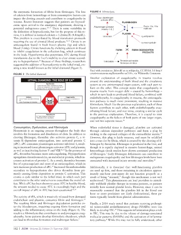
The activity of tPA, which is massively released from damaged tients typically benefit from platelet administration.
endothelium and plasmin, consumes fibrin and fibrinogen.
37
The resulting fibrin and fibrinogen degradation particles en- Finally, a 2014 study noted that patients receiving prehospi-
ter the bloodstream, impairing ongoing fibrin formation and tal nonsteroidal antiinflammatory drugs (NSAIDs) were less
adding to the inflammatory stress on the body (Figure 4). The likely to develop TIC. This suggests inflammation plays a role
41
result is a fibrinolysis that contributes to and propagates coag- in TIC. This may be due to the release of damage- associated
ulopathy. Some patients develop fibrinolytic shutdown, which molecular patterns (DAMPs) and the activation of inflamma-
can lead to thrombus formation and multiorgan failure. 7 tory pathway. While this may not be fully understood, other
4
112 | JSOM Volume 22, Edition 2 / Summer 2022