
Page 117 - JSOM Spring 2021
P. 117
An Ongoing Series
Cutaneous Leishmaniasis
1
Elena M. Crecelius, MD *; Mark W. Burnett, MD 2
ABSTRACT
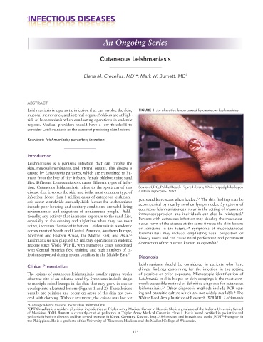
Leishmaniasis is a parasitic infection that can involve the skin, FIGURE 1 An ulcerative lesion caused by cutaneous leishmaniasis.
mucosal membranes, and internal organs. Soldiers are at high-
risk of leishmaniasis when conducting operations in endemic
regions. Medical providers should have a low threshold to
consider Leishmaniasis as the cause of persisting skin lesions.
Keywords: leishmaniasis; parasites; infection
Introduction
Leishmaniasis is a parasitic infection that can involve the
skin, mucosal membranes, and internal organs. This disease is
caused by Leishmania parasites, which are transmitted to hu-
mans from the bite of tiny infected female phlebotomine sand
flies. Different Leishmania spp. cause different types of infec-
tion. Cutaneous leishmaniasis refers to the spectrum of this Source: CDC, Public Health Figure Library, 1962. https://phil.cdc.gov
disease that involves the skin and is the most common type of /Details.aspx?pid=15069
infection. More than 1 million cases of cutaneous leishmani- 1,4
asis occur worldwide annually. Risk factors for leishmaniasis years and leave scars when healed. The skin findings may be
include poor housing and sanitary conditions, crowded living accompanied by nearby swollen lymph nodes. Symptoms of
environments, and migration of nonimmune people. Addi- cutaneous leishmaniasis can recur in the setting of trauma or
1
5
tionally, any activity that increases exposure to the sand flies, immunosuppression and individuals can also be reinfected.
especially in the evening and nighttime when they are most Persons with cutaneous infection may develop the mucocuta-
active, increases the risk of infection. Leishmaniasis is endemic neous form of the disease at the same time as the skin lesions
2,4
across most of South and Central America, Southern Europe, or sometime in the future. Symptoms of mucocutaneous
Northern and Eastern Africa, the Middle East, and Asia. 1,2 leishmaniasis may include long-lasting nasal congestion or
Leishmaniasis has plagued US military operations in endemic bloody noses and can cause nasal perforation and permanent
4
regions since World War II, with numerous cases associated destruction of the mucosa known as espundia.
with Central America field training and high numbers of in-
fections reported during recent conflicts in the Middle East. 3
Diagnosis
Leishmaniasis should be considered in patients who have
Clinical Presentation
clinical findings concerning for the infection in the setting
The lesions of cutaneous leishmaniasis usually appear weeks of possible or prior exposure. Microscopic identification of
after the bite of an infected sand fly. Symptoms include single Leishmania in skin biopsy or skin scrapings is the most com-
to multiple raised bumps in the skin that may grow in size or monly accessible method of definitive diagnosis for cutaneous
develop into ulcerated lesions (Figures 1 and 2). These lesions leishmaniasis. Other diagnostic methods include PCR test-
2,4
usually are painless and occur on areas of the skin not cov- ing and parasitic culture which are not widely available. The
4
ered with clothing. Without treatment, the lesions may last for Walter Reed Army Institute of Research (WRAIR) Leishmania
*Correspondence to elena.m.crecelius.mil@mail.mil
1 CPT Crecelius is a resident physician in pediatrics at Tripler Army Medical Center in Hawaii. She is a graduate of the Indiana University School
of Medicine. COL Burnett is currently chief of pediatrics at Tripler Army Medical Center in Hawaii. He is board certified in pediatrics and
2
pediatric infectious diseases and has served overseas in Korea, Germany, Kosovo, Iraq, Afghanistan, and Kuwait and as the JSOTF-P surgeon in
the Philippines. He is a graduate of the University of Wisconsin-Madison and the Medical College of Wisconsin.
113