
Page 76 - JSOM Summer 2019
P. 76
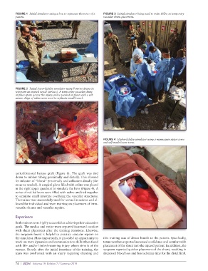
FIGURE 1  Initial simulator using a box to represent the torso of a   FIGURE 3  Initial simulator being used to train 18Ds on temporary
          patient.                                           vascular shunt placement.


          FIGURE 2  Initial lower-fidelity simulator using Penrose drains to
          represent an injured vessel (arrows). A temporary vascular shunt
          in place spans across the injury and is secured in place with a silk
          suture. Bags of saline were used to replicate small bowel.


                                                             FIGURE 4  Higher-fidelity simulator using a mannequin upper torso
                                                             and self-made lower torso.


          aortobifemoral bypass graft (Figure 6). The graft was tied
          down to rubber tubing proximally and distally. This allowed
          for infusion of “blood” proximally and collection distally (for
          reuse as needed). A surgical glove filled with saline was placed
          in the right upper quadrant to simulate the liver (Figure 4). A
          series of red balloons were filled with saline and tied together
          to simulate small intestine overlying the vascular structures.
          The trainer was successfully used for several iterations and al-
          lowed for individual and team training on placement of intra-
          vascular shunts and vascular repairs.


          Experience
          Both trainers were highly successful at achieving their education
          goals. The medics and entire team reported increased comfort
          with shunt placement after the training iterations. Likewise,
          the surgeons found it helpful to practice vascular repairs on
          the simulator. Most importantly, it provided an opportunity to   this training was of direct benefit to the patient. Specifically,
          work on team dynamics and communication skills when faced   team members reported increased confidence and comfort with
          with life- and/or limb-threatening injury where time is of the   placement of the shunt into the injured patient. In addition, the
          essence. Shortly after the initial iterations of the training, the   surgeons reported quicker placement of the shunt, resulting in
          team was confronted with an injury requiring shunting and   decreased blood loss and less ischemia time for the distal limb.


          74  |  JSOM   Volume 19, Edition 2 / Summer 2019