
Page 97 - JSOM Fall 2018
P. 97
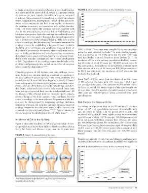
surfaces when the joint moves and assists in joint load bearing. FIGURE 2 Osteoarthritis incidence in the US Military by year.
It is surrounded by synovial fluid, which is contained within
the joint in the joint capsule. Articular cartilage is composed
of a dense, fibrous material (extracellular matrix) that includes
water, collagen fibers, proteoglycans (which fill the spaces be
tween other components and bind them together to maintain
the cartilage structure), and specialized cells called chondro
cytes. The water within the articular cartilage, together with
that in the proteoglycans, is critical for its loadbearing and
lubrication properties. Articular cartilage has no blood vessels,
lymphatics, or nerves and, if damaged, has limited capacity for
repair. Under normal physiological loading of the joint and its
articular cartilage, the chondrocytes function to maintain the
cartilage matrix by establishing a balance between anabolic
(building up or synthesis) and catabolic (breaking down or
degradation) processes in the matrix. However, if an injury oc 2000 to 2015. These rates were compiled from two investiga
8,9
curs or loading of the joint and articular cartilage is excessive, tions that used identical methods. In these studies, spondy
degradation can exceed synthesis, leading to a gradual break losis was defined as OA of the spine and the term “OA” was
down of the articular cartilage and the eventual development used for all other anatomic locations. From 2003 to 2011, the
of OA. Degradation of the cartilage matrix involves the colla incidence of OA in the military population doubled, increas
gen fibers and proteoglycans, as well as a number of enzymes, ing at a rate of about 35 cases per 100,000 personyears. In
which cause the degradation. 10–14 the same period, the incidence of spondylosis increased more
than sixfold at a rate of 69 cases per 100,000 personyears.
Clinical symptoms of OA include joint pain, stiffness, move In 2012 and afterward, the incidence of both disorders has
ment limitations, crepitus (grating, crackling, or creaking in leveled off or declined.
the joint), effusion (excessive fluid in the joint), and bone and
joint deformity. A more definitive diagnosis is usually achieved From 2010 to 2016, more than twothirds of incident cases
from radiographs, by observing jointspace narrowing, osteo of OA involved the knee joint (311 cases per 100,000 per
phytes, sclerosis of the bone covered by the cartilage (subchon sonyears) and shoulders (176 cases per 100,000 personyears).
dral bone). Advanced cases involve subchondral bony cysts In this same period, the lumbar region of the spine was the site
from leakage of synovial fluid into the subchondral bone. All of more than twice the number of incident cases of spondylosis
the tissues of the affected joint are involved (joint capsule, (466 cases per 100,000 person years) compared with other
synovial lining of the joint capsule, bone, cartilage), but the spinal regions.
articular cartilage is the most affected. Progression of the dis
ease can be characterized by decreasing cartilage thickness, Risk Factors for Osteoarthritis
formation of rough and irregular cartilage surfaces, escape of 8,9
cartilage fragments into the joint space, and/or fissures that According to population data in the US military, the inci
can reach into the subchondral bone. 1,11,12,15 Figure 1 shows the dence of OA and spondylosis increased dramatically with
various stages in the development of OA of the knee. 16 age, as shown in Figure 3. In the period 2010 to 2015, the
incidence of OA was 83 times higher among Servicemembers
aged 40 years or older (3,073 cases per 100,000 personyears)
Incidence of OA in the Military when compared with those younger than 20 years (37 cases
Figure 2 shows the incidence of OA and spondylosis in the en per 100,000 person years). For spondylosis, the incidence was
tire population of activeduty Military Servicemembers (Army, 44 times higher among Servicemembers aged 40 years or older
Navy, Air Force, and Marine Corps) over the 16 years from (2,304 cases per 100,000 personyears) when compared with
those younger than 20 years (52 cases per 100,000 person
years). 8
FIGURE 1 Stages of osteoarthritis of the knee.
Besides age, military service, race and ethnicity, and rank were
additional risk factors in the military. Incidence was highest in
FIGURE 3 Osteoarthritis incidence in the US Military by age.
From https://www.medicalmasters.org/kneepaintreatment/
treatingosteoarthritis/
Osteoarthritis and Exercise | 95