
Page 128 - JSOM Winter 2017
P. 128
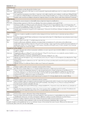
Appendix E  Cont.
           Eyelid laceration
           Goal      Prevent infection, protect the eye from further injury.
           Minimum   Maintain high suspicion for OGI (treat as such if suspected). Keep injured eyelid tissue moist by covering with polyethylene
                     film (food grade).
           Better    For foreign-body penetration, animal bite, or laceration with visible orbital fat, start antibiotic: moxifloxacin 400mg PO daily
                     or levofloxacin 750mg PO daily or amoxicillin/clavulanic acid 875mg/125mg PO every 12 hours or ertapenem 1g IV/IO daily.
           Best      Detailed ocular examination. Irrigate and perform temporary closure of wounds. Tetanus and rabies prophylaxis if indicated.
           Orbital fracture
           Goal      Evaluate for concurrent open or closed globe injury and prevent long-term complications.
           Minimum   Maintain high suspicion for OGI. No nose blowing. Pain control, antiemetic, raise head 30°–45°.
           Better    Antibiotic moxifloxacin 400mg PO daily or levofloxacin 750mg PO daily or amoxicillin/clavulanic acid 875mg/125mg PO
                     every 12 hours or ertapenem 1g IV/IO daily. Initiate nasal decongestant (e.g., Afrin nasal spray twice a day for 3 days) or oral
                     decongestant (pseudoephedrine 30mg every 6 hours).
           Best      Detailed ocular examination. Ice pack for 20 minutes every 1-2 hours for first 48 hours. Monitor for delayed onset of OCS
                     requiring LCC.
           Chemical injuries
           Goal      Initiate eye irrigation as quickly as possible to reduce damage to the eye, treat the injury to minimize scarring and loss of
                     vision.
           Minimum   Immediate irrigation with IV fluid, sterile water, or clean water with at least 2L of fluid. Remove any particulate matter using a
                     cotton tip applicator.
           Better    Continue irrigation until pH = 7, verified using urine test strip.
           Best      Grade I: erythromycin ophthalmic ointment, cycloplegic drops (cyclopentolate 1%), lubrication with artificial tears
                     Grades II–IV: topical antibiotic drops (moxifloxacin 0.5% eye drops, 1 drop every 8 hours), topical corticosteroid (tobradex or
                     prednisolone acetate 1%, 1 drop every hour while awake), doxycycline 100 mg PO every 12 hours, vitamin C 2g 4 times per
                     day, 100% oxygen for 1 hour twice daily
           Preseptal and orbital cellulitis
           Goal      Recognize infection early and start appropriate antibiotics; evacuate suspected cases of orbital cellulitis to an eye surgeon as
                     rapidly as possible
           Minimum   Preseptal: Moxifloxacin 400mg PO daily or levofloxacin 750mg PO daily. Does not cover methicillin-resistant Staphylococcus
                     aureus (MRSA); follow closely for worsening condition
                     Orbital: IV antibiotics: ertapenem, 1g IV/IO daily or levofloxacin 500mg IV once a day
           Better    Orbital: Add nasal decongestant (e.g., Afrin nasal spray twice a day for 3 days) or oral decongestant (pseudoephedrine 30mg
                     every 6 hours)
           Best      Preseptal: Trimethoprim sulfamethoxazole DS 1 tablet PO every 8 hours combined with amoxicillin/clavulanic acid 875mg
                     every 12 hours
                     Orbital: Continue IV antibiotics. Monitor vision every 4 hours until evacuation.
           Infectious keratitis
           Goal      Prompt recognition and treatment to minimize scarring and loss of vision
           Minimum   Moxifloxacin eye drops 1 drop every 15 minutes for first 2 hours, then 1 drop every hour while awake
           Better    Obtain a culture before beginning treatment for sight-threatening keratitis; intense loading dose of moxifloxacin 0.5%
                     eye drops 1 drop every 5–15 minutes for the first 30–60 minutes (patient can self-administer loading dose if reliable) after
                     culture obtained; then 1 drop every 30–60 minutes around the clock until epithelial defect is closed; cycloplegic eye drop
                     (cyclopentolate 1%), 1 drop every 8 hours for photophobia.
           Best      Collagen corneal shield soaked in moxifloxacin drops for transport (5–10 drops) placed over the corneal infiltrate
           Angle-closure glaucoma
           Goal      Prompt recognition and treatment to decrease intraocular pressure
           Minimum   Diagnose based on signs and symptoms: pain, decreased vision, photophobia, dull or cloudy cornea, fixed mid-dilated pupil,
                     increased IOP by palpation. Acetazolamide 500 mg PO initial dose, then 250mg PO every 4 hours to decrease IOP (Note:
                     contrain dicated in patients with sickle cell trait).
           Better    Oral acetazolamide plus topical IOP-lowering eye drops (timolol 0.5%, 1 drop twice a day in the affected eye), antiemetic as
                     needed
           Best      Topical corticosteroid (prednisolone acetate 1%) 1 drop every hour after consultation with eye specialist. IV medication for
                     refractory cases (3% hypertonic saline 250mL IV or mannitol 1g/kg over 30–60 minutes)
           Multitrauma/thermal burn
           Goal      Prevent ocular exposure and corneal injury in high-risk patients.
           Minimum   Keep the ocular surface from drying out by using lubricants: sterile petrolatum or methylcellulose drops (do not substitute a
                     nonophthalmic lubricant). For burns, erythromycin ophthalmic ointment or sterile petrolatum every 2 to 4 hours
           Better    Horizontal taping of eyelids to protect eyes. Evaluate the eyes and instill a lubricant every 8 hours.
           Best      Conduct a detailed ocular examination and cover eyes with food-grade polyethylene film to protect eyes.
           Surgilube should never be instilled into the eye as a lubricant, because of the potential for corneal toxicity. When used for ultrasound
           examination, place a thin film over the closed eyelid.


          126  |  JSOM   Volume 17, Edition 4/Winter 2017