
Page 80 - Journal of Special Operations Medicine - Fall 2017
P. 80
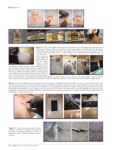
Figure 12  Cont.

                               (C)


           (D)


                                       Figure 13  Foley catheter. (A) Recently, the focus of extended care has brought to the forefront the need
           (A)
                                       for placement of Foley catheters to accurately measure urine output of critically injured or ill patients.
                                       It is possible to have the medic perform a Foley catheter insertion by securing an endotracheal (ET) tube
                                       to the upper thigh (typically with tape or elastic bandage) of the role player so the top lumen is near pu-
                                       bic bone and bot-
                                       tom is at the top   (B)
                                       of thigh. (B) If the
                                       medic determines
                                       that the patient
                                       requires a Foley
                                       catheter, he or she
          can simulate inserting it using a sterile technique by treat-
          ing the bottom of the ET tube along the medial thigh as
          if it were the penis. The catheter should be inserted so the
          balloon goes distal to the superior lumen, then the balloon
          can be inflated and the catheter taped into place. While the
          medic is performing other tasks, or has moved between pa-
          tients, an instructor can occasionally inject liquid with the appropriate shade of yellow into the catheter so the “urine output” can be measured.
          If the scenario involves a patient with crush syndrome or rhabdomyolysis, the instructor can use dark soda in lieu of lighter-colored liquids.

          Figure 14  Chest tube. (A) Demonstrate the incision line on role player, then (B) perform the procedure on a box covered with neoprene. In this
          case, the incision is anterior of where it would be desired and feedback can be given, and the correction could be made. This can be a stand-alone
          anatomy drill where the medic can mark various landmarks on a training partner. If the patient requires a thoracostomy or chest tube, the medic
          should draw an incision line on the role player with a felt-tip marker. If in haste, a chest tube can be secured by multiple chest seals, duct tape,
          and staples, or by suturing. If unable to make the model, taping the chest tube to the patient’s chest emulates what the patient will look like with
          another tube coming out of the body.

                 (A)                              (B)


          Figure 15  Practice suturing on pig feet. If un-
          available, an instructor can make an incision in
          the center of duct-taped neoprene or cloth to a
          table on four sides to allow the student to practice
          suturing.


          78  |  JSOM   Volume 17, Edition 3/Fall 2017