
Page 38 - Journal of Special Operations Medicine - Fall 2017
P. 38
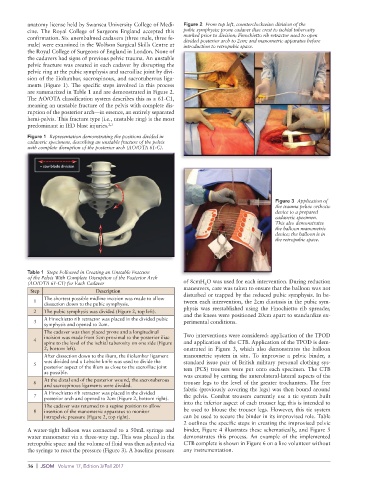
anatomy license held by Swansea University College of Medi-  Figure 2  From top left, counterclockwise: division of the
          cine. The Royal College of Surgeons England accepted this   pubic symphysis; prone cadaver iliac crest to ischial tuberosity
          confirmation. Six unembalmed cadavers (three male, three fe-  marked prior to division; Finochietto rib retractor used to open
          male) were examined in the Wolfson Surgical Skills Centre at   divided posterior arch to 2cm; and manometric apparatus before
                                                             introduction to retropubic space.
          the Royal College of Surgeons of England in London. None of
          the cadavers had signs of previous pelvic trauma. An unstable
          pelvic fracture was created in each cadaver by disrupting the
          pelvic ring at the pubic symphysis and sacroiliac joint by divi-
          sion of the iliolumbar, sacrospinous, and sacrotuberous liga-
          ments (Figure 1). The specific steps involved in this process
          are summarized in Table 1 and are demonstrated in Figure 2.
          The AO/OTA classification system describes this as a 61-C1,
          meaning an unstable fracture of the pelvis with complete dis-
          ruption of the posterior arch—in essence, an entirely separated
          hemi-pelvis. This fracture type (i.e., unstable ring) is the most
          predominant in IED blast injuries. 2,3
          Figure 1  Representation demonstrating the positions divided in
          cadaveric specimens, describing an unstable fracture of the pelvis
          with complete disruption of the posterior arch (AO/OTA 61-C).


                                                                                           Figure 3  Application of
                                                                                           the trauma pelvic orthotic
                                                                                           device to a prepared
                                                                                           cadaveric specimen.
                                                                                           This also demonstrates
                                                                                           the balloon manometric
                                                                                           device; the balloon is in
                                                                                           the retropubic space.


          Table 1  Steps Followed in Creating an Unstable Fracture
          of the Pelvis With Complete Disruption of the Posterior Arch
          (AO/OTA 61-C1) for Each Cadaver                    of 8cmH O was used for each intervention. During reduction
                                                                    2
           Step                 Description                  maneuvers, care was taken to ensure that the balloon was not
                                                             disturbed or trapped by the reduced pubic symphysis. In be-
               The shortest possible midline incision was made to allow
            1                                                tween each intervention, the 2cm diastasis in the pubic sym-
               dissection down to the pubic symphysis.
            2  The pubic symphysis was divided (Figure 2, top left).  physis was reestablished using the Finochietto rib spreader,
                                                             and the knees were positioned 20cm apart to standardize ex-
               A Finochietto rib retractor was placed in the divided pubic
            3                                                perimental conditions.
               symphysis and opened to 2cm.
               The cadaver was then placed prone and a longitudinal
               incision was made from 5cm proximal to the posterior iliac   Two interventions were considered: application of the TPOD
            4  spine to the level of the ischial tuberosity on one side (Figure   and application of the CTB. Application of the TPOD is dem-
               2, bottom left).                              onstrated in Figure 3, which also demonstrates the balloon
               After dissection down to the ilium, the iliolumbar ligament   manometric system in situ. To improvise a pelvic binder, a
               was divided and a Lebsche knife was used to divide the   standard issue pair of British military personal clothing sys-
            5
               posterior aspect of the ilium as close to the sacroiliac joint   tem (PCS) trousers were put onto each specimen. The CTB
               as possible.                                  was created by cutting the anterolateral-lateral aspects of the
               At the distal end of the posterior wound, the sacrotuberous
            6                                                trouser legs to the level of the greater trochanters. The free
               and sacrospinous ligaments were divided.      fabric (previously covering the legs) was then bound around
            7  A Finochietto rib retractor was placed in the divided   the pelvis. Combat trousers currently use a tie system built
               posterior arch and opened to 2cm (Figure 2, bottom right).  into the inferior aspect of each trouser leg; this is intended to
               The cadaver was returned to a supine position to allow
            8  insertion of the manometric apparatus to monitor   be used to blouse the trouser legs. However, this tie system
               intrapelvic pressure (Figure 2, top right).   can be used to secure the binder in its improvised role. Table
                                                             2 outlines the specific steps in creating the improvised pelvic
          A water-tight balloon was connected to a 50mL syringe and   binder, Figure 4 illustrates these schematically, and Figure 5
          water manometer via a three-way tap. This was placed in the   demonstrates this process. An example of the implemented
          retropubic space and the volume of fluid was then adjusted via   CTB complete is shown in Figure 6 on a live volunteer without
          the syringe to reset the pressure (Figure 3). A baseline pressure   any instrumentation.

          36  |  JSOM   Volume 17, Edition 3/Fall 2017