
Page 107 - Journal of Special Operations Medicine - Fall 2014
P. 107
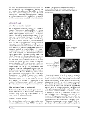
The initial management should be an appropriate his-  Figure 1  Computed tomography scan demonstrating
              tory and physical exam, adequate pain management,   renal calculi (yellow) at uterovesicular junction. There is
              and a clinical determination of the stability of the pa-  significant hydronephrosis as seen by the dilated pelvis of
              tient. In the deployed setting, consider point-of-care ad-  the right kidney.
              juncts such as a rapid urine pregnancy test or a urine dip
              to confirm presence of white blood cells or nitrates. Up
              to 20% of cases of renal calculi will not have hematuria. 3


              KEY QUESTIONS
              Can I clinically make the diagnosis?
              Yes, the diagnosis can be made clinically with reasonable
              certainty. Although there can be variability in presenta-
              tion, the acute onset of unilateral flank pain and hema-
              turia is highly suggestive of renal calculi. The diagnosis
              becomes more certain with the absence of fever, a prior
              history, or positive family history of renal calculi. The
              key question to be asked in the field or forward setting
              would be, What other diagnosis could this be? In the ab-
              sence of pregnancy, peritonitis, and blunt or penetrating
              trauma, the most lethal diagnosis to consider would be
              a ruptured abdominal aortic aneurysm. An abdominal                             Figure 2
              aortic aneurysm is usually a dilation of the aorta, which                      Normal ultrasound
              progresses gradually in some individuals with a history                        appearance of the
              of hypertension. A rupture or leak of this aneurysm can                        right kidney.
              cause severe flank pain, and most emergency physicians
              can cite a case where this presentation can mimic renal
              calculi. These tend to be slow growing and progress over
              years. It is rare that they will rupture or leak if they are
              less than 6cm. Abdominal aortic aneurysms are more
              common after the age of 60 years in men.  A more com-
                                                 4
                                                                             Figure 3
              mon consideration would be the presence of a renal stone   Ultrasound appearance
              and infection that obstructs the ureter. This is known as   of hydronephrosis of
              a septic stone. This can be a true time-sensitive emer-  the right kidney.
              gency, and the patient can become hypotensive and sep-
              tic. This requires emergent drainage to allow drainage of
              the urine around the stone. Fortunately, the clinical his-
              tory, examination, as well as his age and stability make
              these diagnoses very unlikely. In addition, these patholo-  While NSAIDs appear to be about equal to opiates in
              gies can be essential rule out with the use of point-of-care   terms of adequacy of pain control at the 1-hour mark,
              ultrasound (Figures 1–3). As this technology becomes   studies are limited in quality and typically compared with
              more portable, and put into the hands of the tactical   subtherapeutic doses of opiates. Some studies seem to in-
              clinician. Rapid assessment can quickly rule out an ab-  dicate a synergistic role may exist for the combination of
              dominal aortic aneurysm or significant hydronephrosis. 5  these drugs in the treatment of acute renal calculi.  For the
                                                                                                          6
                                                                 tactical clinician, choice of analgesic may be limited based
              What are the risk factors for renal calculi?       on their scope of practice, medication availability, and
                                                                 what opiates they are authorized to use. It would be quite
              Well-documented risk factors include prior history of   reasonable to combine an NSAID and an opiate. Caution
              stones, family history of stones, warmer temperatures,   is suggested in using NSAIDs unless the patient is ade-
              inadequate  fluid  intake,  and  increased  intake  of  cal-  quately hydrated. Giving ibuprofen 400mg orally or ke-
              cium, vitamin C, and animal protein. 5
                                                                 torolac 15–30mg intramuscular (IM)/intravenous (IV) are
                                                                 reasonable choices. Opiate medication could be fentanyl
              How can I treat this acutely?
                                                                 or morphine according to appropriate clinical guidelines.
              The mainstay of treatment for renal calculi has been non-  While some physicians may to want to run IV fluids ag-
              steroidal anti-inflammatory drugs (NSAIDs) and opiates.   gressively to help pass the stone, forced IV therapy is not


              Flank Pain                                                                                      99