
Page 379 - ATP-P 11th Ed
P. 379
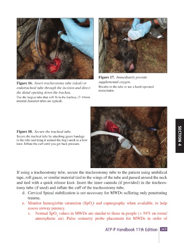
Figure 17.  Immediately provide
        Figure 16.  Insert tracheostomy tube (ideal) or   supplemental oxygen.
        endotracheal tube through the incision and direct   Breathe in the tube or use a hand-operated
        the distal opening down the trachea.  resuscitator.
        Use the largest tube that will fit in the trachea; (7–11mm
        internal diameter tubes are typical).


        Figure 18.  Secure the tracheal tube.
        Secure the tracheal tube by attaching gauze bandage                   SECTION 4
        to the tube and tying it around the dog’s neck in a bow
        knot. Inflate the cuff until you get back pressure.


        If using a tracheostomy tube, secure the tracheostomy tube to the patient using umbilical
        tape, roll gauze, or similar material tied to the wings of the tube and passed around the neck
        and tied with a quick release knot. Insert the inner cannula (if provided) in the tracheos-
        tomy tube (if used) and inflate the cuff of the tracheostomy tube.
           d.  Cervical Spinal stabilization is not necessary for MWDs suffering only penetrating
             trauma.
           e.  Monitor hemoglobin saturation (SpO ) and capnography when available, to help
                                        2
             assess airway patency.
             i.  Normal SpO  values in MWDs are similar to those in people (> 94% on room/
                         2
                atmospheric air). Pulse oximetry probe placement for MWDs in order of
                                              ATP-P Handbook 11th Edition 369